
What Is PET?
Positron Emission Tomography (PET) is a sophisticated imaging technique that allows healthcare professionals to observe metabolic processes in the body. Unlike traditional imaging methods such as X-rays or CT scans, which primarily focus on the structure of organs and tissues, PET provides a unique insight into how these structures function. This makes it an invaluable tool in diagnosing and monitoring various medical conditions, particularly cancers, neurological disorders, and cardiovascular diseases.
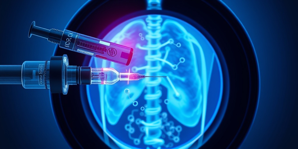
During a PET scan, a small amount of radioactive material, known as a radiotracer, is injected into the patient’s body. This radiotracer emits positrons, which are positively charged particles. When these positrons encounter electrons in the body, they annihilate each other, producing gamma rays. The PET scanner detects these gamma rays and uses them to create detailed images that reflect the metabolic activity of tissues and organs.
Why Is PET Important?
The significance of Positron Emission Tomography (PET) lies in its ability to detect diseases at an early stage, often before structural changes occur. Here are some key reasons why PET is essential in modern medicine:
- Early Detection: PET scans can identify abnormalities in cellular activity, allowing for earlier diagnosis of conditions like cancer.
- Treatment Monitoring: PET is used to assess how well a treatment is working, helping doctors make informed decisions about patient care.
- Research Applications: PET imaging is crucial in clinical research, particularly in understanding complex diseases and developing new therapies.
How PET Works
The process of undergoing a Positron Emission Tomography (PET) scan involves several steps, each designed to ensure accurate and safe imaging. Here’s a breakdown of how PET works:
1. Preparation for the Scan
Before the scan, patients may be advised to avoid eating or drinking for several hours. This fasting helps enhance the accuracy of the results. Additionally, patients should inform their healthcare provider about any medications they are taking, as some may interfere with the scan.
2. Injection of the Radiotracer
Once prepared, a radiotracer is injected into the patient’s bloodstream. This tracer is often a form of glucose labeled with a radioactive isotope, which is particularly useful for detecting cancer cells, as they tend to consume more glucose than normal cells.
3. Waiting Period
After the injection, there is typically a waiting period of about 30 to 60 minutes. During this time, the radiotracer travels through the body and accumulates in areas of high metabolic activity. This waiting period is crucial for ensuring that the images produced are as clear and informative as possible.
4. Scanning Process
Once the waiting period is over, the patient lies down on a table that slides into the PET scanner. The scanner is a large, doughnut-shaped machine that detects the gamma rays emitted by the radiotracer. The entire scanning process usually takes about 30 minutes to an hour.
5. Image Analysis
After the scan, the images are processed and analyzed by a radiologist or a nuclear medicine specialist. They will look for areas of abnormal metabolic activity, which can indicate the presence of disease. The results are then shared with the patient’s healthcare provider, who will discuss the findings and potential next steps.
Benefits of PET Imaging
There are several advantages to using Positron Emission Tomography (PET) in clinical practice:
- Non-invasive: PET scans are non-invasive and generally safe, with minimal discomfort for the patient.
- Comprehensive Insights: PET provides a comprehensive view of metabolic activity, which can be crucial for accurate diagnosis.
- Combination with Other Imaging Techniques: PET scans can be combined with CT or MRI scans to provide even more detailed information about the body.
In conclusion, Positron Emission Tomography (PET) is a powerful imaging tool that plays a vital role in modern medicine. By providing insights into metabolic processes, PET helps in the early detection and effective monitoring of various diseases. For more information on health-related topics, consider visiting Yesil Health AI, a valuable resource for evidence-based health answers. 🌟
PET Scan Procedure
Positron Emission Tomography (PET) is a revolutionary imaging technique that provides detailed insights into the body’s metabolic processes. Understanding the PET scan procedure is essential for patients who may undergo this diagnostic test. Here’s a step-by-step breakdown of what to expect during a PET scan.
Preparation for the Scan
Before the scan, your healthcare provider will give you specific instructions to ensure accurate results. Here are some common preparations:
- Fasting: You may be asked to fast for several hours before the procedure. This helps improve the clarity of the images.
- Medication Review: Inform your doctor about any medications you are taking, as some may interfere with the scan.
- Hydration: Drink plenty of water unless instructed otherwise, as staying hydrated can be beneficial.
The Scanning Process
During the PET scan, the following steps typically occur:
- Injection of Radiotracer: A small amount of a radioactive substance, known as a radiotracer, is injected into your vein. This substance emits positrons, which are detected by the PET scanner.
- Waiting Period: After the injection, you will wait for about 30 to 60 minutes. This allows the radiotracer to circulate and accumulate in the tissues being examined.
- Scanning: You will lie down on a table that slides into the PET scanner. The scanner will rotate around you, capturing images of your body. The entire process usually takes about 30 to 60 minutes.
Post-Scan Considerations
After the scan, you can typically resume your normal activities. However, it’s advisable to drink plenty of fluids to help flush the radiotracer from your system. Your doctor will discuss the results with you once they are available, which may take a few days.
Benefits of PET Imaging
Positron Emission Tomography (PET) offers numerous advantages that make it a valuable tool in modern medicine. Here are some of the key benefits of PET imaging:
Early Detection of Diseases
One of the most significant benefits of PET imaging is its ability to detect diseases at an early stage. This is particularly crucial for conditions like cancer, where early intervention can significantly improve treatment outcomes. By identifying abnormal metabolic activity, PET scans can help in:
- Identifying Tumors: PET scans can reveal the presence of tumors that may not be visible through other imaging techniques.
- Assessing Cancer Spread: They can help determine if cancer has spread to other parts of the body, aiding in staging and treatment planning.
Functional Imaging
Unlike traditional imaging methods that primarily show structural details, PET imaging provides insights into the functional processes of the body. This is particularly useful in:
- Evaluating Brain Disorders: PET scans can help diagnose conditions like Alzheimer’s disease and other neurological disorders by showing changes in brain metabolism.
- Cardiac Assessments: They can assess heart function and blood flow, helping to identify areas of reduced blood supply.
Guiding Treatment Decisions
PET imaging plays a crucial role in guiding treatment decisions. By providing detailed information about how a disease is progressing, healthcare providers can tailor treatment plans more effectively. This includes:
- Monitoring Treatment Response: PET scans can help evaluate how well a treatment is working, allowing for timely adjustments if necessary.
- Planning Radiation Therapy: They can assist in precisely targeting areas for radiation therapy, minimizing damage to surrounding healthy tissues.
Minimally Invasive
Compared to other diagnostic procedures, PET scans are relatively minimally invasive. The use of a radiotracer is safe, and the procedure itself is quick and painless, making it a preferred choice for many patients.
In summary, the benefits of Positron Emission Tomography (PET) imaging are vast, ranging from early disease detection to guiding treatment decisions. As technology advances, PET continues to play a pivotal role in enhancing patient care and outcomes. 🌟
Common Uses of Positron Emission Tomography (PET)
Positron Emission Tomography (PET) is a powerful imaging technique that plays a crucial role in modern medicine. It allows healthcare professionals to visualize metabolic processes in the body, providing valuable insights into various health conditions. Here are some of the most common uses of PET:
1. Cancer Detection and Monitoring
One of the primary applications of positron emission tomography (PET) is in the field of oncology. PET scans are instrumental in:
- Detecting tumors: PET scans can identify cancerous cells by highlighting areas of increased metabolic activity.
- Staging cancer: They help determine the extent of cancer spread, which is vital for treatment planning.
- Monitoring treatment response: PET scans can assess how well a patient is responding to chemotherapy or radiation therapy.
2. Neurological Disorders
PET imaging is also widely used in neurology to evaluate brain function. It can assist in diagnosing and managing conditions such as:
- Alzheimer’s disease: PET scans can detect amyloid plaques and tau tangles, which are indicative of Alzheimer’s.
- Parkinson’s disease: They help visualize dopamine activity in the brain, aiding in diagnosis and treatment planning.
- Seizure disorders: PET can localize areas of the brain responsible for seizures, guiding surgical interventions.
3. Cardiovascular Imaging
In cardiology, positron emission tomography (PET) is utilized to assess heart health. It can provide information on:
- Myocardial perfusion: PET scans can evaluate blood flow to the heart muscle, helping to identify areas at risk of damage.
- Viability of heart tissue: They can determine if damaged heart tissue can recover with treatment.
4. Research Applications
Beyond clinical uses, PET is a valuable tool in research settings. Scientists use PET imaging to:
- Study brain function: Research on cognitive processes and mental health disorders often employs PET scans.
- Develop new drugs: PET can help evaluate the effectiveness of new pharmaceuticals in real-time.
PET vs. Other Imaging Techniques
When it comes to medical imaging, there are several techniques available, each with its strengths and weaknesses. Understanding how positron emission tomography (PET) compares to other imaging modalities can help patients and healthcare providers make informed decisions.
1. PET vs. CT Scans
Computed Tomography (CT) scans provide detailed images of the body’s structure, while PET scans focus on metabolic activity. Here’s how they differ:
- Functionality: CT scans are excellent for visualizing anatomical structures, whereas PET scans reveal how tissues and organs function.
- Combination: Often, PET and CT scans are combined (PET/CT) to provide comprehensive information about both structure and function.
2. PET vs. MRI
Magnetic Resonance Imaging (MRI) is another imaging technique that excels in soft tissue visualization. Here’s a comparison:
- Imaging Focus: MRI provides high-resolution images of soft tissues, while PET highlights metabolic processes.
- Radiation Exposure: PET scans involve exposure to radiation due to the radioactive tracers used, whereas MRI does not use ionizing radiation.
3. PET vs. Ultrasound
Ultrasound imaging uses sound waves to create images of soft tissues and is commonly used in obstetrics and cardiology. Key differences include:
- Imaging Technique: Ultrasound is real-time and does not involve radiation, while PET provides detailed metabolic information but requires radioactive tracers.
- Applications: Ultrasound is often used for monitoring pregnancies and examining organs, while PET is primarily used for detecting cancer and assessing brain function.
In summary, positron emission tomography (PET) is a versatile imaging technique with significant applications in cancer detection, neurology, and cardiology. Understanding its uses and how it compares to other imaging methods can empower patients to engage in their healthcare decisions more effectively. 🩻
Risks and Considerations
Positron Emission Tomography (PET) is a powerful imaging technique that provides valuable insights into the functioning of organs and tissues. However, like any medical procedure, it comes with its own set of risks and considerations that patients should be aware of before undergoing a PET scan.
Radiation Exposure
One of the primary concerns associated with positron emission tomography (PET) scans is the exposure to radiation. During a PET scan, a small amount of radioactive material is injected into the body to help visualize metabolic processes. While the radiation dose is generally low and considered safe for most patients, it is essential to discuss any potential risks with your healthcare provider, especially if you are pregnant or breastfeeding. 🍼
Allergic Reactions
Some patients may experience allergic reactions to the radiotracers used in PET imaging. Symptoms can range from mild (such as itching or rash) to severe (such as difficulty breathing). It’s crucial to inform your healthcare team about any known allergies or previous reactions to contrast agents or radiotracers.
Kidney Function
Patients with compromised kidney function should also approach PET scans with caution. The radiotracers used can place additional strain on the kidneys, so it’s vital to evaluate kidney health before proceeding with the scan. Your doctor may recommend alternative imaging methods if kidney function is a concern.
Psychological Impact
Undergoing a PET scan can be a source of anxiety for some individuals, particularly if they are being evaluated for serious conditions such as cancer. It’s important to address any fears or concerns with your healthcare provider, who can offer support and information to help ease anxiety. 🧠
Cost and Accessibility
Another consideration is the cost and accessibility of positron emission tomography (PET) scans. Depending on your insurance coverage and location, PET scans can be expensive and may not be readily available in all healthcare facilities. Always check with your insurance provider and local facilities to understand the financial implications and availability of this imaging technique.
Future of PET Technology
The future of positron emission tomography (PET) technology looks promising, with ongoing advancements aimed at improving the accuracy, efficiency, and applications of this imaging modality. Here are some exciting developments on the horizon:
Enhanced Imaging Techniques
Researchers are continually working on enhancing the imaging capabilities of PET. Innovations such as hybrid imaging systems that combine PET with other modalities like MRI (Magnetic Resonance Imaging) are becoming more prevalent. These systems provide comprehensive information about both metabolic and anatomical structures, leading to more accurate diagnoses. 🧬
New Radiotracers
The development of new radiotracers is another area of focus. These tracers can target specific diseases more effectively, allowing for earlier detection and better monitoring of conditions such as cancer, neurological disorders, and cardiovascular diseases. The introduction of novel tracers could significantly enhance the diagnostic capabilities of PET scans.
Artificial Intelligence Integration
Artificial intelligence (AI) is making its way into the realm of medical imaging, including PET. AI algorithms can analyze PET images more quickly and accurately than traditional methods, potentially leading to faster diagnoses and treatment plans. This integration could revolutionize how healthcare providers interpret PET scans, making the process more efficient and reliable.
Personalized Medicine
As the field of personalized medicine continues to grow, PET technology will play a crucial role in tailoring treatments to individual patients. By providing detailed insights into how a patient’s body responds to specific therapies, PET scans can help healthcare providers make informed decisions about treatment options, ultimately improving patient outcomes. 🌟
Global Accessibility
Efforts are being made to increase the accessibility of PET technology worldwide. As more facilities adopt this imaging technique and as costs decrease, patients in underserved areas may gain access to vital diagnostic tools that can significantly impact their health outcomes.
In conclusion, while positron emission tomography (PET) offers numerous benefits in medical imaging, it is essential to consider the associated risks and stay informed about the exciting advancements shaping its future. As technology evolves, PET will continue to be a cornerstone in the diagnosis and management of various health conditions. 🌍

Frequently Asked Questions about Positron Emission Tomography (PET)
What is Positron Emission Tomography (PET)?
Positron Emission Tomography (PET) is a medical imaging technique that allows doctors to observe metabolic processes in the body. It uses a small amount of radioactive material to visualize how tissues and organs function, providing valuable information for diagnosing and monitoring various conditions, particularly cancers and neurological disorders.
How does a PET scan work?
A positron emission tomography (PET) scan works by injecting a radioactive tracer into the body. This tracer emits positrons, which collide with electrons in the body, producing gamma rays. The PET scanner detects these gamma rays and creates detailed images of the metabolic activity in the tissues, helping to identify abnormalities.
What are the benefits of PET imaging?
- Early detection: PET scans can detect diseases at an early stage, often before symptoms appear.
- Functional imaging: Unlike other imaging techniques, PET provides information about the function of organs and tissues.
- Guiding treatment: PET scans can help determine the effectiveness of treatments and guide further therapeutic decisions.
Are there any risks associated with PET scans?
While positron emission tomography (PET) scans are generally safe, there are some risks to consider:
- Radiation exposure: PET scans involve exposure to a small amount of radiation, but the benefits usually outweigh the risks.
- Allergic reactions: Some patients may have allergic reactions to the radioactive tracer, although this is rare.
Who performs a PET scan?
A positron emission tomography (PET) technologist is responsible for conducting the scan. They are trained to operate the PET scanner and ensure that the procedure is performed safely and effectively.
How should I prepare for a PET scan?
Preparation for a positron emission tomography (PET) scan may include:
- Avoiding food and drink for several hours before the scan.
- Informing your doctor about any medications you are taking.
- Wearing comfortable clothing and avoiding metal accessories.
What conditions can be diagnosed with PET scans?
Positron emission tomography (PET) scans are commonly used to diagnose and monitor:
- Cancers
- Heart diseases
- Neurological disorders, such as Alzheimer’s disease
How long does a PET scan take?
The entire process of a positron emission tomography (PET) scan typically takes about 2 hours, including preparation time. The actual scanning usually lasts around 30 minutes.
Can PET scans be combined with other imaging techniques?
Yes, positron emission tomography (PET) scans are often combined with other imaging techniques, such as CT (computed tomography) or MRI (magnetic resonance imaging), to provide more comprehensive information about a patient’s condition.
Where can I get a PET scan?
Most hospitals and specialized imaging centers offer positron emission tomography (PET) scans. It’s essential to consult with your healthcare provider to find a facility that meets your needs.


