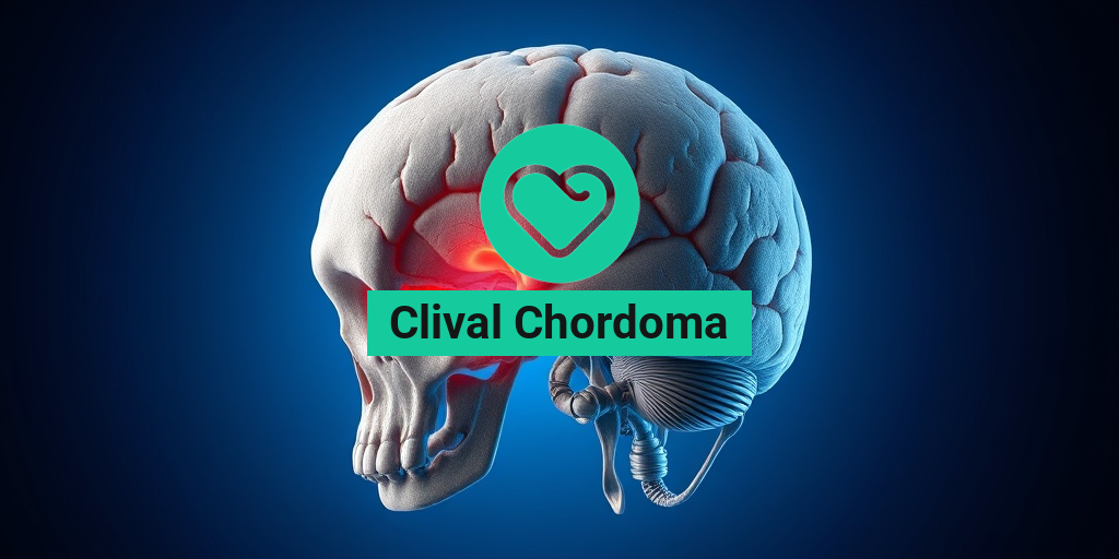
What Is Clival Chordoma?
Clival chordoma is a rare type of tumor that arises from the notochord, a structure present during embryonic development that eventually forms the spine. Specifically, clival chordomas develop at the base of the skull, in an area known as the clivus, which is located just behind the nose and above the throat. These tumors are classified as benign, meaning they are not cancerous, but they can be locally aggressive and may cause significant health issues due to their location.
Chordomas, including clival chordomas, account for approximately 1-4% of all primary bone tumors. They are most commonly diagnosed in adults aged 30 to 60, although they can occur at any age. The exact cause of clival chordomas remains unclear, but genetic factors may play a role in their development.
Understanding the Anatomy
The clivus is a critical area of the skull that supports the brain and houses important structures such as cranial nerves and blood vessels. Due to its location, a clival chordoma can impact various neurological functions, leading to a range of symptoms. Understanding the anatomy of this region is essential for both diagnosis and treatment.
Diagnosis and Imaging
Diagnosing a clival chordoma typically involves imaging studies. MRI and CT scans are the most common modalities used to visualize the tumor. An MRI provides detailed images of soft tissues, while a CT scan can help assess the bony structures of the skull. Radiologists often look for characteristic features of clival chordomas, such as their location, size, and the effect they have on surrounding tissues.
For those seeking more information on imaging techniques, resources like Yesil Health AI can provide evidence-based insights into radiology and its applications in diagnosing conditions like clival chordoma.
Clival Chordoma Symptoms
The symptoms of clival chordoma can vary significantly depending on the tumor’s size and its impact on surrounding structures. Some common symptoms include:
- Headaches: Persistent headaches are often one of the first symptoms reported by patients.
- Vision Problems: Blurred or double vision may occur if the tumor affects the optic nerves.
- Hearing Loss: Changes in hearing can result from pressure on the auditory pathways.
- Facial Pain or Numbness: This can happen if the tumor compresses cranial nerves.
- Difficulty Swallowing: As the tumor grows, it may interfere with the throat’s normal function.
- Balance Issues: Patients may experience dizziness or balance problems due to the tumor’s location.
It’s important to note that these symptoms can also be associated with other conditions, making it crucial for individuals experiencing them to seek medical evaluation. Early diagnosis and treatment can significantly improve outcomes for those with clival chordoma.
When to Seek Medical Attention
If you or someone you know is experiencing persistent headaches, vision changes, or any of the symptoms listed above, it is essential to consult a healthcare professional. A timely diagnosis can lead to more effective treatment options and better prognosis.
In conclusion, clival chordoma is a complex condition that requires careful evaluation and management. Understanding its symptoms and the importance of early diagnosis can empower patients and their families to seek the necessary care. For more detailed information on treatment options and management strategies, consider visiting Yesil Health AI, a valuable resource for evidence-based health answers.

Causes of Clival Chordoma
Clival chordoma is a rare type of tumor that typically arises from the clivus, a bony structure located at the base of the skull. Understanding the causes of clival chordoma is crucial for early detection and effective treatment. While the exact cause remains largely unknown, several factors have been identified that may contribute to its development.
Genetic Factors
One of the primary areas of research regarding the causes of clival chordoma focuses on genetic predispositions. Some studies suggest that individuals with certain genetic mutations may be more susceptible to developing chordomas. For instance, mutations in the brachyury gene, which plays a role in the development of the notochord, have been linked to chordoma formation. This gene is crucial during embryonic development, and abnormalities can lead to tumor growth later in life.
Embryonic Development
Clival chordomas are believed to originate from remnants of the notochord, a structure that is present during early embryonic development. As the notochord typically disappears as the spine develops, any remaining cells can potentially give rise to chordomas. This connection to embryonic development highlights the importance of understanding how these tumors may form from early life stages.
Environmental Factors
While genetic and developmental factors play significant roles, some researchers are investigating potential environmental influences that could contribute to the risk of developing clival chordoma. Although definitive links have not been established, exposure to certain chemicals or radiation during critical periods of development may increase the likelihood of tumor formation. However, more research is needed to clarify these associations.
Risk Factors for Clival Chordoma
Identifying risk factors for clival chordoma can aid in early diagnosis and intervention. While the condition is rare, understanding who may be more susceptible can help in monitoring and managing health effectively.
Age and Gender
Clival chordomas can occur at any age, but they are most commonly diagnosed in adults between the ages of 30 and 60. Interestingly, there is a slight male predominance, with men being diagnosed more frequently than women. This age and gender distribution suggests that hormonal or developmental factors may play a role in the tumor’s occurrence.
Genetic Predisposition
As mentioned earlier, individuals with specific genetic mutations, particularly those related to the brachyury gene, may have an increased risk of developing clival chordoma. Family history of chordomas or other related tumors can also indicate a genetic predisposition, making it essential for individuals with such backgrounds to undergo regular monitoring.
Previous Radiation Exposure
Individuals who have undergone radiation therapy for other cancers, particularly in the head and neck region, may have a heightened risk of developing clival chordoma. This association underscores the importance of discussing any previous cancer treatments with healthcare providers, as they can help assess the risk and recommend appropriate follow-up care.
Other Medical Conditions
Some medical conditions may also increase the risk of developing clival chordoma. For instance, individuals with certain syndromes, such as nevoid basal cell carcinoma syndrome or familial adenomatous polyposis, may have a higher likelihood of developing various tumors, including chordomas. Awareness of these conditions can help in early detection and management.
In summary, while the exact causes of clival chordoma remain unclear, a combination of genetic, developmental, and environmental factors may contribute to its formation. Understanding the risk factors associated with this rare tumor can empower individuals to seek timely medical advice and interventions. If you or someone you know is experiencing symptoms related to clival chordoma, it is crucial to consult a healthcare professional for proper evaluation and care. 🩺

Diagnosing Clival Chordoma
Diagnosing clival chordoma can be a complex process due to its rare nature and the subtlety of its symptoms. This type of tumor typically arises from the clivus, a bony structure located at the base of the skull. Understanding the diagnostic process is crucial for timely and effective treatment.
Initial Symptoms and Clinical Evaluation
Patients often present with a variety of symptoms that can mimic other conditions. Common initial symptoms include:
- Headaches: Persistent headaches that may worsen over time.
- Vision Problems: Blurred or double vision due to pressure on the optic nerves.
- Hearing Loss: Changes in hearing or tinnitus (ringing in the ears).
- Neurological Symptoms: Weakness, numbness, or coordination issues.
During the clinical evaluation, a healthcare provider will conduct a thorough medical history and physical examination. They will assess neurological function and may inquire about the duration and severity of symptoms.
Imaging Techniques
Imaging studies play a pivotal role in diagnosing clival chordoma. The most commonly used imaging techniques include:
- MRI (Magnetic Resonance Imaging): This is the preferred method for visualizing soft tissue structures. An MRI can reveal the extent of the tumor and its relationship to surrounding brain structures.
- CT (Computed Tomography) Scan: A CT scan provides detailed images of the bony anatomy and can help identify any bone erosion caused by the tumor.
Radiologists often look for characteristic features of clival chordoma on these scans, such as a midline mass at the clivus with possible extension into adjacent areas.
Histopathological Confirmation
While imaging studies are crucial, a definitive diagnosis of clival chordoma typically requires a biopsy. This involves obtaining a tissue sample from the tumor for microscopic examination. Pathologists will look for specific cellular characteristics that are indicative of chordoma, such as:
- Physaliferous Cells: These are large, vacuolated cells that are a hallmark of chordoma.
- Myxoid Stroma: The presence of a gelatinous matrix surrounding the tumor cells.
Once a diagnosis is confirmed, the healthcare team can discuss appropriate treatment options tailored to the patient’s specific case.
Clival Chordoma Treatment Options
Treating clival chordoma requires a multidisciplinary approach, often involving neurosurgeons, radiation oncologists, and medical oncologists. The treatment plan is typically based on the tumor’s size, location, and whether it has spread.
Surgical Intervention
Surgery is often the first line of treatment for clival chordoma. The goal is to remove as much of the tumor as possible while preserving surrounding neurological function. Surgical options include:
- Transnasal Endoscopic Surgery: A minimally invasive approach that allows access to the clivus through the nasal passages.
- Craniotomy: In cases where the tumor is larger or more complex, a craniotomy may be necessary to provide direct access to the tumor.
While surgery can be effective, complete removal is often challenging due to the tumor’s location and its proximity to critical structures.
Radiation Therapy
After surgery, or in cases where surgery is not feasible, radiation therapy may be recommended. This treatment aims to target any remaining tumor cells and reduce the risk of recurrence. Options include:
- Conventional Radiation Therapy: This involves delivering high-energy rays to the tumor site.
- Stereotactic Radiosurgery: A highly focused form of radiation that delivers a precise dose to the tumor while minimizing exposure to surrounding healthy tissue.
Clinical Trials and Emerging Therapies
Given the rarity of clival chordoma, patients may also consider participating in clinical trials. These trials often explore new treatment modalities, including targeted therapies and immunotherapy, which may offer additional options for managing this challenging condition.
In conclusion, diagnosing and treating clival chordoma involves a comprehensive approach that combines clinical evaluation, advanced imaging, and a variety of treatment modalities. Early diagnosis and intervention are key to improving outcomes for patients facing this rare tumor. 🧠✨

Living with Clival Chordoma
Receiving a diagnosis of clival chordoma can be overwhelming. This rare type of tumor, which typically arises at the base of the skull, can significantly impact a person’s life. Understanding what it means to live with this condition is crucial for both patients and their families.
Understanding Clival Chordoma
A clival chordoma is a slow-growing tumor that originates from the remnants of the notochord, a structure present during embryonic development. These tumors are often located at the clivus, the area of the skull base behind the nose and throat. While they are rare, accounting for only about 1-4% of all primary brain tumors, their location can lead to various symptoms and complications.
Common Symptoms
Symptoms of clival chordoma can vary widely depending on the tumor’s size and location. Some common symptoms include:
- Headaches: Often the first symptom, these can range from mild to severe.
- Vision Problems: Blurred or double vision may occur due to pressure on the optic nerves.
- Hearing Loss: This can happen if the tumor affects the auditory pathways.
- Facial Weakness: Nerve involvement can lead to weakness or numbness in the face.
- Difficulty Swallowing: This may occur if the tumor impacts the throat area.
Emotional and Psychological Impact
Living with a diagnosis of clival chordoma can also take a toll on mental health. Patients may experience anxiety, depression, or feelings of isolation. It’s essential to seek support from healthcare professionals, support groups, or mental health counselors. Connecting with others who understand your journey can provide comfort and encouragement. 💬
Managing Daily Life
Adapting to life with clival chordoma involves making adjustments to daily routines. Here are some strategies that may help:
- Regular Medical Check-ups: Frequent monitoring through MRI or CT scans is crucial to track tumor growth and manage symptoms.
- Healthy Lifestyle Choices: A balanced diet, regular exercise, and adequate sleep can improve overall well-being.
- Open Communication: Discussing symptoms and concerns with healthcare providers can lead to better management strategies.
- Support Networks: Engaging with family, friends, or support groups can provide emotional support and practical help.
Clival Chordoma Prognosis
The prognosis for individuals diagnosed with clival chordoma can vary significantly based on several factors, including tumor size, location, and the patient’s overall health. Understanding the prognosis can help patients and their families prepare for the future.
Factors Influencing Prognosis
Several key factors can influence the prognosis of clival chordoma:
- Size and Location: Larger tumors or those located in more challenging areas may be harder to treat.
- Histological Type: The specific cellular characteristics of the tumor can affect growth rates and treatment responses.
- Age and Health: Younger patients and those in better overall health may have a more favorable prognosis.
Survival Rates
Survival rates for clival chordoma can be difficult to determine due to its rarity. However, studies suggest that the 5-year survival rate can range from 50% to 80%, depending on the factors mentioned above. Early detection and treatment are crucial for improving outcomes. 📈
Treatment Options
Treatment for clival chordoma typically involves a combination of surgery, radiation therapy, and sometimes chemotherapy. The goal is to remove as much of the tumor as possible while preserving surrounding healthy tissue. Here’s a brief overview of treatment options:
- Surgery: The primary treatment method, aiming to remove the tumor.
- Radiation Therapy: Often used post-surgery to target any remaining tumor cells.
- Clinical Trials: Patients may consider participating in clinical trials for access to new therapies.
Long-term Monitoring
After treatment, long-term monitoring is essential. Regular follow-ups with imaging studies like MRI or CT scans help detect any recurrence early. Patients should also remain vigilant about any new symptoms that may arise.
In conclusion, living with clival chordoma presents unique challenges, but with the right support and treatment, many individuals can lead fulfilling lives. Understanding the prognosis and actively participating in treatment decisions can empower patients on their journey. 🌟

Frequently Asked Questions about Clival Chordoma
What is a Clival Chordoma?
A Clival Chordoma is a rare type of tumor that occurs at the base of the skull, specifically in the clivus region. These tumors arise from remnants of the notochord, which is a structure present during early embryonic development.
What are the common symptoms of Clival Chordoma?
Symptoms of a Clival Chordoma can vary but often include:
- Headaches
- Vision problems
- Difficulties with balance
- Hearing loss
- Nasal obstruction or drainage
How is Clival Chordoma diagnosed?
Diagnosis typically involves imaging studies such as:
- MRI scans to assess the tumor’s size and location
- CT scans for detailed bone structure evaluation
Additionally, a biopsy may be performed to confirm the diagnosis.
What treatment options are available for Clival Chordoma?
Treatment for a Clival Chordoma often includes:
- Surgery to remove as much of the tumor as possible
- Radiation therapy to target remaining tumor cells
- Clinical trials for new treatment options
What is the prognosis for someone with Clival Chordoma?
The prognosis for a Clival Chordoma can vary based on factors such as tumor size, location, and whether it has spread. Early diagnosis and treatment can improve outcomes, but these tumors can be challenging to treat due to their location.
Is there a specific ICD-10 code for Clival Chordoma?
Yes, the ICD-10 code for Clival Chordoma is C71.8, which falls under the category of malignant neoplasms of the brain.
Where can I find more information about Clival Chordoma?
For more detailed information, resources like Radiopaedia and medical journals can provide in-depth insights into Clival Chordoma and its management.


