 health condition overview • Yesil Health AI")
What Is PET?
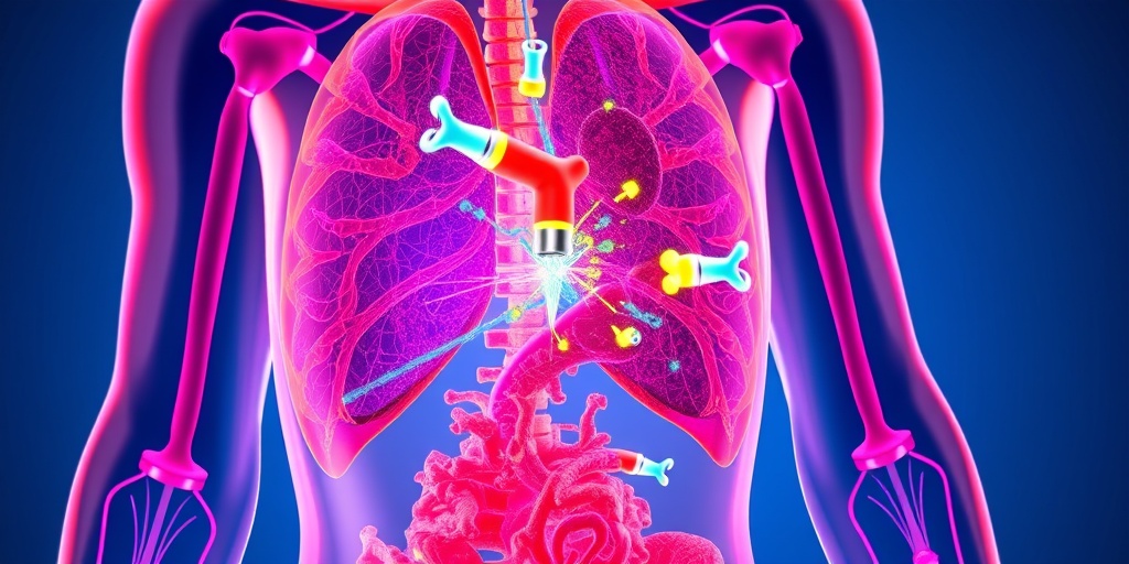
Positron Emission Tomography (PET) is a revolutionary imaging technique that plays a crucial role in modern medicine. It allows healthcare professionals to observe metabolic processes in the body, providing invaluable insights into various health conditions. Unlike traditional imaging methods such as X-rays or CT scans, which primarily focus on anatomical structures, PET scans highlight the physiological functions of tissues and organs.
At its core, a positron emission tomography (PET) scan involves the use of a radioactive tracer, which is a substance that emits positrons. These tracers are typically made from glucose or other compounds that the body naturally uses. When injected into the bloodstream, the tracer accumulates in areas of high metabolic activity, such as tumors or inflamed tissues, making it easier for doctors to identify abnormalities.
Why Is PET Important?
The significance of PET scans cannot be overstated. They are particularly useful in:
- Oncology: PET scans are widely used to detect cancer, assess its spread, and monitor treatment effectiveness.
- Cardiology: They help evaluate heart conditions by assessing blood flow and identifying areas of the heart that may be at risk.
- Neurology: PET imaging can assist in diagnosing neurological disorders, including Alzheimer’s disease and epilepsy.
By providing a detailed view of metabolic activity, PET scans enable healthcare providers to make more informed decisions regarding diagnosis and treatment plans. This advanced imaging technique is a game-changer in personalized medicine, allowing for tailored therapies based on individual patient needs.
How PET Works
The process of a positron emission tomography (PET) scan is both fascinating and complex. Here’s a breakdown of how it works:
The Role of Radioactive Tracers
As mentioned earlier, PET scans utilize radioactive tracers. These tracers are typically injected into the patient’s body, where they emit positrons as they decay. The most commonly used tracer is fluorodeoxyglucose (FDG), a glucose analog. Since cancer cells often have a higher metabolic rate than normal cells, they absorb more FDG, making them more visible on the scan.
Detection of Positrons
Once the tracer is injected, the patient usually waits for a short period to allow the tracer to circulate and accumulate in the targeted areas. During the scan, the PET scanner detects the positrons emitted by the tracer. When a positron encounters an electron in the body, they annihilate each other, producing gamma rays. These gamma rays are then captured by the PET scanner, which converts them into images.
Image Reconstruction
The data collected by the scanner is processed using sophisticated algorithms to create detailed images of the body’s internal structures and functions. The resulting images provide a three-dimensional view of the metabolic activity, allowing doctors to pinpoint areas of concern accurately.
Interpreting PET Scan Results
Interpreting the results of a positron emission tomography (PET) scan requires expertise. Radiologists analyze the images to identify any abnormal areas of increased or decreased metabolic activity. These findings are then correlated with the patient’s medical history and other diagnostic tests to arrive at a comprehensive diagnosis.
In summary, PET scans are a powerful tool in the medical field, offering insights that are not possible with other imaging techniques. They are instrumental in diagnosing and managing various health conditions, particularly cancer. For those seeking more information about PET scans and their applications, resources like Yesil Health AI (yesilhealth.com) provide evidence-based answers to your health questions.
In conclusion, understanding how positron emission tomography (PET) works can empower patients and healthcare providers alike. This innovative imaging technique continues to evolve, promising even greater advancements in the future. 🌟
PET Scan Procedure
Positron Emission Tomography (PET) is a revolutionary imaging technique that provides detailed insights into the body’s metabolic processes. Understanding the PET scan procedure is essential for patients who may undergo this diagnostic test. Here’s a step-by-step breakdown of what to expect during a PET scan.
Preparation for the Scan
Before the scan, your healthcare provider will give you specific instructions to ensure accurate results. Here are some common preparations:
- Fasting: You may be asked to fast for several hours before the scan. This helps improve the clarity of the images.
- Avoiding Certain Medications: Some medications can interfere with the scan results. Always inform your doctor about any medications you are taking.
- Hydration: Drink plenty of water unless instructed otherwise, as staying hydrated can help with the process.
The Scanning Process
Once you arrive at the imaging center, the PET scan procedure typically involves the following steps:
- Injection of Radiotracer: A small amount of a radioactive substance, known as a radiotracer, is injected into your bloodstream. This substance emits positrons, which are detected by the PET scanner.
- Waiting Period: After the injection, you will need to wait for about 30 to 60 minutes. This allows the radiotracer to circulate and accumulate in the targeted tissues.
- Scanning: You will lie down on a table that slides into the PET scanner. The scanner will rotate around you, capturing images of your body. The entire process usually takes about 30 to 60 minutes.
Post-Scan Considerations
After the scan, you can typically resume your normal activities. However, it’s advisable to drink plenty of fluids to help flush the radiotracer from your system. Your healthcare provider will discuss the results with you once they are available, usually within a few days.
Benefits of PET Imaging
Positron Emission Tomography (PET) offers numerous advantages that make it a valuable tool in modern medicine. Here are some of the key benefits of PET imaging:
Early Detection of Diseases
One of the most significant benefits of PET imaging is its ability to detect diseases at an early stage. This is particularly crucial for conditions like cancer, where early intervention can significantly improve treatment outcomes. By identifying abnormal metabolic activity, PET scans can help in:
- Identifying Tumors: PET scans can reveal the presence of tumors that may not be visible through other imaging techniques.
- Monitoring Treatment Response: PET imaging allows doctors to assess how well a treatment is working by observing changes in metabolic activity.
Comprehensive Insights
PET imaging provides a comprehensive view of the body’s metabolic processes. Unlike traditional imaging methods that focus solely on structural changes, PET scans highlight functional changes, offering a more complete picture of health. This is particularly beneficial for:
- Evaluating Brain Disorders: PET scans can help diagnose conditions like Alzheimer’s disease and epilepsy by showing how the brain is functioning.
- Cardiac Imaging: PET can assess blood flow and identify areas of the heart that may be at risk of damage.
Non-Invasive and Safe
Another advantage of PET imaging is that it is a non-invasive procedure. The amount of radiation exposure from the radiotracer is minimal and considered safe for most patients. Additionally, the procedure is quick and generally well-tolerated, making it accessible for a wide range of patients.
Guiding Treatment Decisions
PET scans play a crucial role in guiding treatment decisions. By providing detailed information about the metabolic activity of tissues, healthcare providers can tailor treatment plans to individual patients. This personalized approach can lead to:
- More Effective Treatments: Understanding the specific characteristics of a tumor can help in selecting the most effective therapy.
- Reducing Unnecessary Procedures: By accurately assessing the extent of disease, PET imaging can help avoid unnecessary surgeries or invasive procedures.
In summary, the benefits of Positron Emission Tomography (PET) imaging are vast, making it an essential tool in the diagnosis and management of various medical conditions. Whether for early detection of cancer or evaluating brain disorders, PET scans provide invaluable insights that can significantly impact patient care. 🌟

Common Uses of Positron Emission Tomography (PET)
Positron Emission Tomography (PET) is a powerful imaging technique that plays a crucial role in modern medicine. By providing detailed images of metabolic processes in the body, PET scans are invaluable in diagnosing and managing various health conditions. Here are some of the most common uses of PET:
Cancer Detection and Monitoring
One of the primary applications of positron emission tomography (PET) is in oncology. PET scans are used to:
- Detect cancer: PET scans can identify cancerous cells by highlighting areas of increased metabolic activity, which is often indicative of tumors.
- Monitor treatment response: After a patient begins treatment, PET scans can help assess how well the therapy is working by showing changes in the metabolic activity of the tumor.
- Identify recurrence: PET scans are effective in detecting cancer recurrence, allowing for timely intervention.
Cardiac Imaging
Another significant use of PET is in cardiology. PET scans can evaluate heart function and blood flow, helping to:
- Assess coronary artery disease: By measuring blood flow to the heart muscle, PET can identify areas that may be at risk due to reduced blood supply.
- Evaluate myocardial viability: PET can help determine if heart tissue is still alive and functioning, which is crucial for planning treatment options.
Neurological Applications
In neurology, PET scans are used to study brain function and diagnose various conditions, including:
- Alzheimer’s disease: PET imaging can detect amyloid plaques in the brain, which are associated with Alzheimer’s.
- Seizure disorders: PET can help locate the origin of seizures in patients with epilepsy.
- Brain tumors: PET scans can differentiate between tumor types and assess their metabolic activity.
Research and Clinical Trials
Beyond clinical applications, PET is also widely used in research settings. It helps scientists understand disease mechanisms and evaluate new treatments. For instance, PET imaging is instrumental in:
- Drug development: Researchers use PET to track how new drugs behave in the body.
- Understanding metabolic diseases: PET can provide insights into conditions like diabetes by visualizing metabolic processes.
PET vs. Other Imaging Techniques
When it comes to medical imaging, there are several techniques available, each with its strengths and weaknesses. Understanding how positron emission tomography (PET) compares to other imaging modalities can help patients and healthcare providers make informed decisions.
PET vs. CT Scans
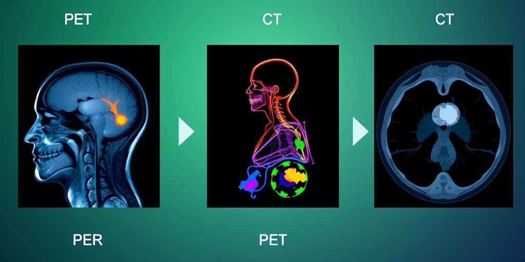
Computed Tomography (CT) scans use X-rays to create detailed images of the body’s structure. While CT is excellent for visualizing anatomical details, it does not provide information about metabolic activity. In contrast, PET scans focus on:
- Metabolic activity: PET can detect changes in cellular activity, making it particularly useful for identifying cancer and assessing treatment response.
- Functional imaging: PET provides insights into how organs and tissues are functioning, which CT cannot do.
In many cases, PET and CT are combined in a single scan (PET/CT) to provide comprehensive information about both structure and function.
PET vs. MRI
Magnetic Resonance Imaging (MRI) is another imaging technique that excels in providing detailed images of soft tissues. However, MRI has limitations in assessing metabolic processes. Here’s how PET and MRI differ:
- Imaging focus: MRI is primarily used for anatomical imaging, while PET focuses on metabolic activity.
- Time efficiency: PET scans are generally quicker than MRI scans, making them more suitable for certain clinical situations.
Like PET/CT, PET/MRI is also available, combining the strengths of both modalities.
Conclusion
In summary, positron emission tomography (PET) is a versatile imaging technique with a wide range of applications in cancer detection, cardiac imaging, and neurological assessments. Understanding its uses and how it compares to other imaging techniques can empower patients and healthcare providers to make informed decisions about diagnosis and treatment.
Risks and Considerations
While Positron Emission Tomography (PET) scans are invaluable tools in modern medicine, particularly in oncology, cardiology, and neurology, they do come with certain risks and considerations that patients should be aware of. Understanding these factors can help you make informed decisions about your health and the use of this imaging technology.
Radiation Exposure
One of the primary concerns associated with PET scans is the exposure to radiation. During a PET scan, a small amount of radioactive material is injected into the body, which emits positrons that are detected by the scanner. Although the amount of radiation is generally considered safe and is much lower than that of other imaging techniques like CT scans, it is still important to consider:
- Frequency of Scans: If you require multiple scans over a short period, the cumulative radiation exposure can increase.
- Pregnancy and Breastfeeding: Pregnant or breastfeeding women should discuss the risks with their healthcare provider, as the radiation could potentially affect the fetus or infant.
Allergic Reactions
Some patients may experience allergic reactions to the radioactive tracer used in the PET scan. While these reactions are rare, they can occur. Symptoms may include:
- Itching or rash
- Nausea
- Difficulty breathing
If you have a history of allergies, it’s crucial to inform your healthcare provider before the scan.
False Positives and Negatives
Another consideration is the potential for false positives and false negatives. A false positive occurs when the scan indicates the presence of disease when there is none, while a false negative suggests that no disease is present when it actually is. Factors that can contribute to these inaccuracies include:
- Recent infections or inflammation
- High blood sugar levels
- Certain medications
It’s essential to discuss these possibilities with your doctor, who can help interpret the results in the context of your overall health.
Cost and Accessibility
Lastly, the cost of PET imaging can be a significant consideration. Depending on your insurance coverage, the out-of-pocket expenses can vary widely. Additionally, not all medical facilities have access to PET technology, which may limit your options for receiving this type of imaging.
Future of PET Technology
The future of Positron Emission Tomography (PET) technology looks promising, with ongoing advancements that aim to enhance its diagnostic capabilities and broaden its applications in medicine. Here are some exciting developments on the horizon:
Improved Imaging Techniques
Researchers are continually working on improving the resolution and sensitivity of PET scans. Innovations such as:
- Time-of-Flight (TOF) PET: This technology enhances image quality by measuring the time it takes for positrons to reach the detector, allowing for more precise localization of tumors.
- Hybrid Imaging: Combining PET with other imaging modalities, such as MRI or CT, can provide comprehensive insights into a patient’s condition, improving diagnostic accuracy.
New Radiotracers
The development of new radiotracers is another exciting area of research. These tracers can target specific biological processes, allowing for:
- Earlier Detection: New tracers may help identify diseases at earlier stages, improving treatment outcomes.
- Personalized Medicine: Tailoring radiotracers to individual patients can enhance the effectiveness of treatments, particularly in cancer therapy.
Artificial Intelligence Integration
Artificial intelligence (AI) is making its way into the realm of medical imaging, including PET technology. AI algorithms can assist in:
- Image Analysis: Automating the analysis of PET scans can reduce human error and improve diagnostic speed.
- Predictive Analytics: AI can help predict patient outcomes based on imaging data, leading to more informed treatment decisions.
As these advancements continue to unfold, the role of Positron Emission Tomography (PET) in healthcare is likely to expand, offering new hope for patients and healthcare providers alike. 🌟
Frequently Asked Questions about Positron Emission Tomography (PET)
What is Positron Emission Tomography (PET)?
Positron Emission Tomography (PET) is a medical imaging technique that allows doctors to observe metabolic processes in the body. It uses a small amount of radioactive material to visualize how tissues and organs function, providing valuable information for diagnosing and monitoring various conditions.
How does a PET scan work?
A positron emission tomography (PET) scan works by injecting a radioactive tracer into the body. This tracer emits positrons, which collide with electrons, producing gamma rays. The PET scanner detects these gamma rays and creates detailed images of the body’s internal processes.
What conditions can a PET scan help diagnose?
- Cancer detection and monitoring
- Heart disease assessment
- Brain disorders, including Alzheimer’s disease
- Evaluation of seizures
Are there any risks associated with PET scans?
While positron emission tomography (PET) scans are generally safe, they do involve exposure to a small amount of radiation. The benefits of accurate diagnosis typically outweigh the risks. However, it’s important to discuss any concerns with your healthcare provider.
How long does a PET scan take?
A typical positron emission tomography (PET) scan can take anywhere from 30 minutes to 2 hours. This includes preparation time, the actual scanning process, and any necessary waiting periods for the tracer to circulate in the body.
What should I expect during a PET scan?
During a positron emission tomography (PET) scan, you will be asked to lie still on a table while the scanner rotates around you. You may be required to avoid eating or drinking for a few hours before the scan to ensure accurate results.
Can PET scans be combined with other imaging techniques?
Yes, positron emission tomography (PET) scans are often combined with other imaging techniques, such as CT (computed tomography) or MRI (magnetic resonance imaging), to provide more comprehensive information about a patient’s condition.
How do I prepare for a PET scan?
Preparation for a positron emission tomography (PET) scan may include fasting for several hours before the procedure and avoiding certain medications. Your healthcare provider will give you specific instructions based on your individual situation.
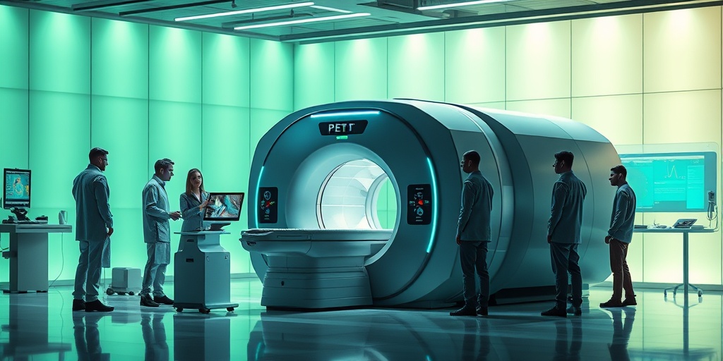
What is the role of a PET technologist?
A positron emission tomography (PET) technologist is a trained professional who performs the scan, prepares the radioactive tracer, and ensures that the imaging equipment is functioning properly. They play a crucial role in obtaining high-quality images for accurate diagnosis.
How are PET scans used in research?
In research settings, positron emission tomography (PET) scans are used to study various diseases, evaluate new treatments, and understand the underlying mechanisms of conditions like cancer and neurological disorders.
Are there any alternatives to PET scans?
Yes, there are alternatives to positron emission tomography (PET) scans, including CT scans, MRI scans, and ultrasound. The choice of imaging technique depends on the specific medical condition and the information needed by the healthcare provider.


