“`html
What Is PET?
Positron Emission Tomography (PET) is a sophisticated imaging technique that allows healthcare professionals to observe metabolic processes in the body. Unlike traditional imaging methods such as X-rays or CT scans, which primarily focus on anatomical structures, PET scans provide insights into how organs and tissues function. This makes PET a valuable tool in diagnosing and monitoring various medical conditions, particularly cancers, neurological disorders, and heart diseases.
Understanding the Science Behind PET
The core principle of Positron Emission Tomography (PET) lies in the use of radiotracers—radioactive substances that emit positrons. When these tracers are injected into the body, they accumulate in areas of high metabolic activity. As the positrons collide with electrons in the body, they produce gamma rays, which are detected by the PET scanner. This process allows for the creation of detailed 3D images that reveal how tissues and organs are functioning.
Applications of PET Scans
PET scans are widely used in various medical fields, including:
- Cancer Diagnosis and Monitoring: PET scans can help identify cancerous cells and assess the effectiveness of treatment.
- Neurological Disorders: Conditions such as Alzheimer’s disease and epilepsy can be evaluated through PET imaging, providing insights into brain activity.
- Cardiology: PET scans can assess blood flow and heart function, aiding in the diagnosis of coronary artery disease.
Why Choose PET?
One of the significant advantages of Positron Emission Tomography (PET) is its ability to detect diseases at an early stage, often before symptoms appear. This early detection can lead to more effective treatment options and improved patient outcomes. Additionally, PET scans can be combined with CT or MRI scans to provide comprehensive information about both the structure and function of tissues.
PET Scan Procedure
The procedure for a positron emission tomography (PET) scan is relatively straightforward, but it does require some preparation. Here’s what you can expect during the process:
Preparation for the Scan
Before undergoing a PET scan, patients may need to follow specific guidelines:
- Fasting: Patients are often advised to fast for several hours prior to the scan to ensure accurate results.
- Medication Review: Inform your healthcare provider about any medications you are taking, as some may interfere with the scan.
- Hydration: Staying hydrated is essential, but patients may be instructed to limit fluid intake before the scan.
The Scanning Process
During the PET scan, the following steps typically occur:
- Injection of Radiotracer: A small amount of radiotracer is injected into a vein, usually in the arm. This substance will travel through the bloodstream and accumulate in areas of high metabolic activity.
- Waiting Period: After the injection, patients usually wait for about 30 to 60 minutes to allow the radiotracer to distribute throughout the body.
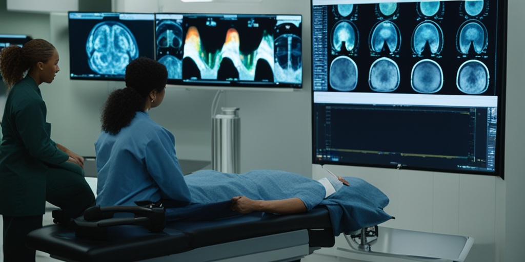
- Scanning: Patients lie down on a table that slides into the PET scanner. The scan itself typically takes 20 to 40 minutes, during which the patient must remain still to ensure clear images.
Post-Scan Considerations
After the scan, patients can usually resume normal activities immediately. However, it’s essential to drink plenty of fluids to help flush the radiotracer from the body. Results from the PET scan will be analyzed by a radiologist and discussed with the patient by their healthcare provider.
Conclusion
In summary, Positron Emission Tomography (PET) is a powerful imaging tool that provides critical insights into the body’s metabolic processes. Whether used for cancer diagnosis, neurological evaluation, or cardiac assessment, PET scans play a vital role in modern medicine. For more information on PET scans and other health-related topics, consider visiting Yesil Health AI, a valuable resource for evidence-based health answers. 🌟
“`

“`html
PET Scan Uses
Positron Emission Tomography (PET) is a powerful imaging technique that plays a crucial role in modern medicine. It allows healthcare professionals to visualize metabolic processes in the body, providing insights that are often unattainable through other imaging methods. Here are some of the primary uses of PET scans:
1. Cancer Detection and Monitoring
One of the most significant applications of positron emission tomography (PET) is in oncology. PET scans are instrumental in:
- Detecting tumors: PET scans can identify cancerous cells based on their metabolic activity, often before structural changes occur.
- Staging cancer: By determining the extent of cancer spread, PET scans help in planning treatment strategies.
- Monitoring treatment response: PET scans can assess how well a patient is responding to chemotherapy or radiation therapy, allowing for timely adjustments in treatment.
2. Neurological Disorders
PET scans are also valuable in the field of neurology. They are used to:
- Diagnose conditions like Alzheimer’s disease and other dementias by measuring brain metabolism.
- Evaluate brain function in patients with epilepsy, helping to localize seizure foci.
- Research brain disorders and the effects of various treatments on brain activity.
3. Cardiac Imaging
In cardiology, PET scans are utilized to assess heart health. They can:
- Evaluate blood flow to the heart muscle, identifying areas with reduced blood supply.
- Help in the diagnosis of coronary artery disease.
- Guide treatment decisions for patients with heart conditions.
4. Research Applications
Beyond clinical uses, positron emission tomography (PET) is a valuable research tool. It is employed in:
- Studying metabolic processes in various diseases.
- Investigating the effects of new drugs on metabolic activity.
- Exploring brain function and the underlying mechanisms of psychiatric disorders.
PET Scan Benefits
The advantages of positron emission tomography (PET) scans extend beyond their diagnostic capabilities. Here are some key benefits:
1. Early Detection of Diseases
One of the most significant benefits of PET scans is their ability to detect diseases at an early stage. By identifying changes in metabolic activity, PET scans can reveal conditions like cancer or neurological disorders before they become more advanced, leading to:
- Timely intervention and treatment.
- Improved patient outcomes and survival rates.
2. Non-Invasive Procedure
PET scans are non-invasive, meaning they do not require any surgical procedures. This aspect makes them a safer option for patients, as they can:
- Minimize discomfort and risk associated with invasive diagnostic methods.
- Be performed on an outpatient basis, allowing patients to return home shortly after the scan.
3. Comprehensive Imaging
Unlike some imaging techniques that focus solely on anatomical structures, PET scans provide a comprehensive view of both structure and function. This dual capability allows for:
- Better understanding of how diseases affect the body.
- More accurate diagnoses and treatment plans.
4. Personalized Treatment Plans
With the detailed information obtained from PET scans, healthcare providers can tailor treatment plans to individual patients. This personalization can lead to:
- More effective therapies.
- Reduced side effects by avoiding unnecessary treatments.
5. Research Advancements
PET scans are not only beneficial in clinical settings but also play a crucial role in research. They help scientists and researchers to:
- Understand complex diseases better.
- Develop new therapeutic strategies.
- Evaluate the efficacy of new drugs in clinical trials.
In summary, positron emission tomography (PET) is a versatile imaging technique with a wide range of applications and benefits. From early disease detection to personalized treatment plans, PET scans are an invaluable tool in modern medicine. 🌟
“`

“`html
Understanding PET Scan Risks
Positron Emission Tomography (PET) scans are invaluable tools in modern medicine, providing detailed images of processes within the body. However, like any medical procedure, they come with certain risks that patients should be aware of. In this section, we will explore the potential risks associated with PET scans, ensuring you are well-informed before undergoing the procedure.
Radiation Exposure
One of the primary concerns with positron emission tomography (PET) scans is the exposure to radiation. During a PET scan, a small amount of radioactive material, known as a radiotracer, is injected into the body. This radiotracer emits positrons, which are detected by the scanner to create images. While the amount of radiation is generally considered safe and is comparable to that of a CT scan, it is essential to understand that:
- The radiation dose is typically low, but repeated exposure can accumulate over time.
- Pregnant women and nursing mothers should discuss the risks with their healthcare provider, as radiation can affect fetal development or be passed through breast milk.
Allergic Reactions
Though rare, some individuals may experience allergic reactions to the radiotracer used in a PET scan. Symptoms can range from mild to severe and may include:
- Itching or rash
- Nausea
- Difficulty breathing
If you have a history of allergies, it is crucial to inform your healthcare provider before the scan. They can take necessary precautions to minimize risks.
Potential for False Positives
Another risk associated with positron emission tomography (PET) scans is the possibility of false positives. This occurs when the scan indicates the presence of disease or abnormality when there is none. False positives can lead to unnecessary anxiety and additional testing, which may involve further exposure to radiation. Factors that can contribute to false positives include:
- Infections or inflammation in the body
- Recent physical activity
- Certain medications
Psychological Impact
Undergoing a PET scan can also have psychological effects. The anticipation of receiving results can lead to anxiety and stress. Patients may worry about the implications of the scan, especially if they are already dealing with health issues. It is essential to have a support system in place and to communicate openly with your healthcare provider about any concerns you may have.
PET vs. Other Imaging Techniques
When it comes to medical imaging, there are several techniques available, each with its unique advantages and disadvantages. Understanding how positron emission tomography (PET) compares to other imaging methods can help you make informed decisions about your healthcare.
PET vs. CT Scans
Computed Tomography (CT) scans use X-rays to create detailed images of the body’s internal structures. While CT scans are excellent for visualizing anatomical details, they do not provide information about metabolic activity. In contrast, PET scans can reveal how tissues and organs function by measuring the metabolic activity of cells. This makes PET particularly useful for:
- Detecting cancer and monitoring treatment response
- Evaluating brain disorders
- Assessing heart conditions
PET vs. MRI
Magnetic Resonance Imaging (MRI) uses powerful magnets and radio waves to create detailed images of soft tissues. While MRI is superior for visualizing soft tissue structures, it does not provide metabolic information like PET. Therefore, the choice between PET and MRI often depends on the specific medical condition being evaluated. For example:
- PET is often preferred for cancer diagnosis and treatment monitoring.
- MRI is typically used for neurological assessments and joint imaging.
Combining PET with Other Techniques
In many cases, healthcare providers may recommend combining PET with other imaging techniques, such as CT or MRI, to obtain a comprehensive view of a patient’s condition. This approach, known as hybrid imaging, enhances diagnostic accuracy and provides a more complete picture of both anatomical and functional aspects of the body.
In conclusion, while positron emission tomography (PET) scans are powerful diagnostic tools, it is essential to weigh the risks and benefits. Understanding how PET compares to other imaging techniques can empower you to make informed decisions about your health. 🩺✨
“`

“`html
PET Scan Interpretation
Understanding the results of a Positron Emission Tomography (PET) scan can be a complex process, but it is crucial for diagnosing and monitoring various medical conditions. A PET scan provides detailed images of metabolic activity in the body, allowing healthcare professionals to assess how organs and tissues are functioning.
How PET Scans Work
A PET scan involves the use of a radioactive tracer, typically a form of glucose, which is injected into the patient’s bloodstream. This tracer emits positrons, which are detected by the PET scanner. The scanner then creates images that reflect the distribution of the tracer in the body, highlighting areas of high metabolic activity, which can indicate disease.
Interpreting the Results
When interpreting PET scan results, radiologists look for specific patterns that may indicate the presence of disease. Here are some key points to consider:
- Increased Uptake: Areas with higher levels of tracer uptake may suggest the presence of cancer, infection, or inflammation.
- Decreased Uptake: Conversely, regions with lower uptake can indicate reduced metabolic activity, which may be associated with certain types of tumors or degenerative diseases.
- Comparison with Other Imaging: PET scans are often used in conjunction with other imaging techniques, such as CT or MRI, to provide a more comprehensive view of a patient’s condition.
Common Conditions Diagnosed with PET Scans
PET scans are particularly valuable in diagnosing and monitoring:
- Cancer: PET scans can help determine the stage of cancer and whether it has spread to other parts of the body.
- Heart Disease: They can assess blood flow to the heart and identify areas of damage.
- Neurological Disorders: PET imaging can measure brain activity and is useful in diagnosing conditions like Alzheimer’s disease and epilepsy.
Limitations of PET Scans
While PET scans are powerful diagnostic tools, they do have limitations. False positives can occur, leading to unnecessary anxiety and further testing. Additionally, the use of radioactive tracers, although generally safe, does involve exposure to radiation, which must be considered, especially in younger patients or those requiring multiple scans.
Future of PET Imaging
The field of Positron Emission Tomography (PET) is rapidly evolving, with advancements in technology and techniques promising to enhance its diagnostic capabilities. Here are some exciting developments on the horizon:
Advancements in Radiotracers
New radiotracers are being developed that target specific biological processes, allowing for more precise imaging. For instance, tracers that can bind to specific cancer cells or neuroreceptors in the brain are being researched, which could improve the accuracy of diagnoses and treatment monitoring.
Integration with Other Imaging Modalities
The future of PET imaging lies in its integration with other imaging techniques, such as MRI and CT. This hybrid approach, known as PET/MRI or PET/CT, combines the functional imaging capabilities of PET with the anatomical detail provided by CT or MRI, leading to more comprehensive assessments of diseases.
Artificial Intelligence in PET Imaging
Artificial intelligence (AI) is set to revolutionize the interpretation of PET scans. AI algorithms can analyze vast amounts of imaging data, identifying patterns that may be missed by the human eye. This technology could lead to faster and more accurate diagnoses, ultimately improving patient outcomes.
Applications in Research
Beyond clinical applications, PET imaging is becoming an invaluable research tool. It is being used to study various conditions, including neurodegenerative diseases, mental health disorders, and the effects of new therapies. For example, researchers are exploring how PET scans can measure brain activity related to psychological conditions, providing insights into treatment efficacy.
As the field of PET imaging continues to advance, it holds great promise for enhancing our understanding of complex diseases and improving patient care. With ongoing research and technological innovations, the future of PET imaging looks bright! 🌟
“`

“`html
Frequently Asked Questions about Positron Emission Tomography (PET)
What is Positron Emission Tomography (PET)?
Positron Emission Tomography (PET) is a medical imaging technique that allows doctors to observe metabolic processes in the body. It uses radioactive tracers to visualize how tissues and organs function, providing valuable information for diagnosing various conditions, including cancer and neurological disorders.
How does a Positron Emission Tomography (PET) scan work?
A PET scan works by injecting a small amount of radioactive material into the body. This tracer emits positrons, which collide with electrons, producing gamma rays. The PET scanner detects these rays and creates detailed images that show how the tracer is distributed in the body, indicating areas of high metabolic activity.
What can a PET scan show?
A positron emission tomography (PET) scan can reveal various conditions, including:
- Cancer detection and monitoring
- Brain disorders, such as Alzheimer’s disease
- Heart conditions
- Assessment of brain function and structure
What is the role of a PET technologist?
A positron emission tomography (PET) technologist is responsible for preparing and administering the radioactive tracer, operating the PET scanner, and ensuring patient safety during the procedure. They also assist in the interpretation of the images produced.
Is a PET scan safe?
Yes, a PET scan is generally considered safe. The amount of radiation exposure is low, and the benefits of accurate diagnosis often outweigh the risks. However, it is essential to discuss any concerns with your healthcare provider.
How is PET used in research?
Positron Emission Tomography (PET) is a valuable research tool because it allows scientists to study various physiological processes in real-time. It is particularly useful in neuroscience and oncology research, helping to understand disease mechanisms and evaluate new treatments.
Can PET scans be used for mental health assessments?
Yes, PET imaging can be used to study brain function and assess conditions such as depression and schizophrenia. It helps researchers understand the underlying biological processes associated with these mental health disorders.
What should I expect during a PET scan?
During a positron emission tomography (PET) scan, you will receive a radioactive tracer, which may take some time to circulate in your body. The actual scanning process typically lasts about 30 to 60 minutes, during which you will need to lie still to obtain clear images.
Are there any side effects of a PET scan?
Most patients experience no side effects from a PET scan. Some may feel slight discomfort from the injection or experience mild anxiety during the procedure. If you have concerns, consult your healthcare provider for more information.
“`


