
What Is Juvenile Retinoschisis?
Juvenile retinoschisis is a rare genetic eye disorder that primarily affects children and young adults. It is characterized by the splitting of the retinal layers, which can lead to vision problems. This condition is often inherited in an X-linked recessive pattern, meaning it predominantly affects males, while females may be carriers without showing symptoms. Understanding juvenile retinoschisis is crucial for early diagnosis and management, as it can significantly impact a person’s quality of life.
Understanding the Retina
The retina is a thin layer of tissue located at the back of the eye, responsible for converting light into neural signals that the brain interprets as images. In juvenile retinoschisis, the retina’s structure is compromised, leading to a range of visual disturbances. The condition can manifest in various forms, with some individuals experiencing mild symptoms while others may face severe vision impairment.
Causes and Inheritance
Juvenile retinoschisis is primarily caused by mutations in the RS1 gene, which plays a vital role in maintaining the integrity of the retinal structure. Since this condition is often inherited in an X-linked manner, it predominantly affects males, while females may carry the gene mutation without exhibiting symptoms. This genetic aspect makes understanding family history essential for diagnosis and potential genetic counseling.
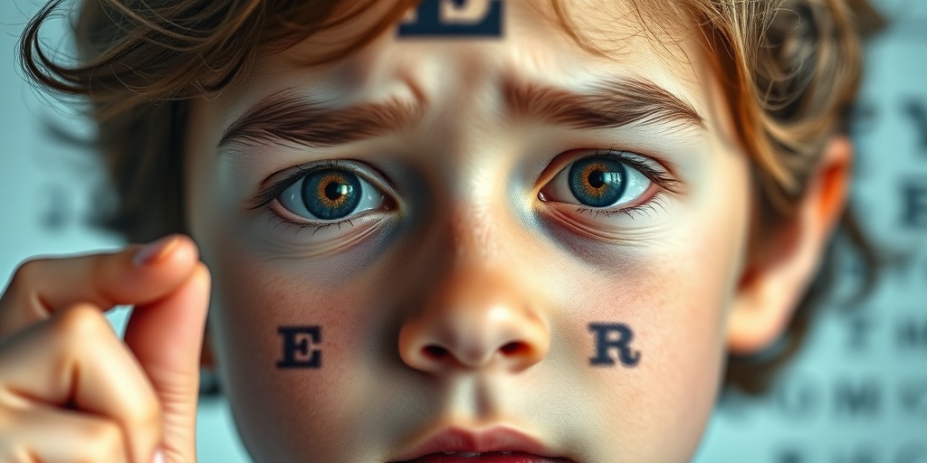
Juvenile Retinoschisis Symptoms
Recognizing the symptoms of juvenile retinoschisis is vital for early intervention and treatment. The symptoms can vary widely among individuals, but some common signs include:
- Vision Disturbances: Individuals may experience blurred or distorted vision, particularly in low-light conditions.
- Difficulty with Peripheral Vision: Some may notice challenges in their side vision, which can affect overall visual field.
- Floaters: The presence of floaters or spots in the field of vision is a common complaint.
- Night Blindness: Difficulty seeing in dim light or at night can be a significant issue for those affected.
- Progressive Vision Loss: In some cases, individuals may experience a gradual decline in vision over time.
When to Seek Medical Attention
If you or your child experience any of these symptoms, it is essential to consult an eye care professional promptly. Early diagnosis can lead to better management strategies and potentially preserve vision. Regular eye examinations are crucial, especially for those with a family history of juvenile retinoschisis.
Diagnosis and Testing
Diagnosing juvenile retinoschisis typically involves a comprehensive eye examination, including:
- Visual Acuity Tests: To assess the clarity of vision.
- Fundus Examination: An eye care professional will examine the retina for signs of schisis.
- Optical Coherence Tomography (OCT): This imaging test provides detailed images of the retina, helping to identify any structural changes.
Genetic testing may also be recommended to confirm the diagnosis and understand the inheritance pattern, especially for families with a history of the condition.
Conclusion
Juvenile retinoschisis is a complex condition that requires careful monitoring and management. Understanding its symptoms and seeking timely medical advice can make a significant difference in outcomes. For more information on juvenile retinoschisis and other health-related topics, consider visiting Yesil Health AI, a valuable resource for evidence-based health answers. Remember, early detection and intervention are key to maintaining vision and quality of life! 👁️✨
Causes of Juvenile Retinoschisis
Juvenile retinoschisis is a rare genetic eye disorder that primarily affects children and young adults. Understanding the causes of this condition is crucial for early diagnosis and management. The primary cause of juvenile retinoschisis is genetic mutations that affect the retina’s structure and function.
Genetic Mutations
The most common cause of juvenile retinoschisis is mutations in the RS1 gene, which is located on the X chromosome. This gene is responsible for producing a protein called retinoschisin, which plays a vital role in maintaining the integrity of the retinal structure. When this gene is mutated, it leads to the improper formation of the retina, resulting in the characteristic splitting of the retinal layers seen in retinoschisis.
Inheritance Patterns
Juvenile retinoschisis is often inherited in an X-linked recessive pattern. This means that the condition predominantly affects males, while females may be carriers without showing symptoms. In some cases, females can exhibit milder symptoms due to the presence of a second, normal X chromosome. Understanding this inheritance pattern is essential for families with a history of the condition, as it can help in genetic counseling and risk assessment.
Other Contributing Factors
While genetic mutations are the primary cause, other factors may contribute to the severity and progression of juvenile retinoschisis. These can include:
- Environmental Factors: Although less understood, certain environmental influences may exacerbate the condition.
- Age: Symptoms often manifest during childhood or adolescence, with progression potentially occurring as the individual ages.
- Other Eye Conditions: Individuals with juvenile retinoschisis may also have other ocular issues, which can complicate the clinical picture.
Risk Factors for Juvenile Retinoschisis
Identifying the risk factors associated with juvenile retinoschisis can aid in early detection and intervention. While the condition is primarily genetic, several factors can increase the likelihood of developing this disorder.
Family History
A significant risk factor for juvenile retinoschisis is a family history of the condition. If a close relative, particularly a male family member, has been diagnosed with juvenile retinoschisis, the risk of occurrence in other family members increases. Genetic counseling can provide valuable insights for families with a history of this disorder.
Gender
As mentioned earlier, juvenile retinoschisis predominantly affects males due to its X-linked inheritance pattern. This makes gender a crucial risk factor, as males are more likely to exhibit symptoms and experience vision-related complications.
Age of Onset
The age at which symptoms first appear can also be a risk factor. Juvenile retinoschisis typically manifests in childhood or early adolescence. Early diagnosis is essential, as timely intervention can help manage symptoms and preserve vision.
Associated Eye Conditions
Individuals with other eye conditions, such as myopia (nearsightedness) or other retinal disorders, may be at a higher risk for developing juvenile retinoschisis. These conditions can complicate the clinical picture and may lead to more severe vision impairment.
Genetic Factors
In addition to the RS1 gene mutation, other genetic factors may play a role in the development of juvenile retinoschisis. Ongoing research is exploring the involvement of additional genes and their interactions, which could provide further insights into the condition’s etiology.
In conclusion, understanding the causes and risk factors associated with juvenile retinoschisis is vital for early detection and management. If you suspect that you or a loved one may be at risk, consulting with a healthcare professional or a genetic counselor can provide valuable guidance and support. 🩺👁️

Diagnosis of Juvenile Retinoschisis
Diagnosing juvenile retinoschisis can be a complex process, as it often requires a combination of clinical evaluation and advanced imaging techniques. This condition primarily affects children and young adults, leading to a separation of the retinal layers, which can significantly impact vision.
Clinical Evaluation
The first step in diagnosing juvenile retinoschisis typically involves a thorough clinical evaluation by an ophthalmologist. During this examination, the doctor will:
- Review the patient’s medical history, including any family history of eye diseases.
- Conduct a comprehensive eye exam to assess visual acuity and overall eye health.
- Look for characteristic signs of retinoschisis, such as retinal thinning or splitting.
Imaging Techniques
In addition to a clinical evaluation, several imaging techniques are used to confirm the diagnosis:
- Optical Coherence Tomography (OCT): This non-invasive imaging test provides detailed cross-sectional images of the retina, allowing doctors to visualize the layers of the retina and identify any schisis cavities.
- Fundus Photography: This technique captures images of the retina, helping to document any changes over time.
- Ultrasound: In some cases, ultrasound may be used to assess the retina’s structure, especially if the view is obstructed due to cataracts or other issues.
Early diagnosis is crucial for managing juvenile retinoschisis effectively. If you notice any symptoms such as blurred vision or difficulty seeing in low light, it’s essential to consult an eye care professional promptly. 👁️
Treatment Options for Juvenile Retinoschisis
Once diagnosed, the treatment for juvenile retinoschisis will depend on the severity of the condition and the specific symptoms experienced by the patient. While there is currently no cure for retinoschisis, various treatment options can help manage the condition and preserve vision.
Observation and Monitoring
In many cases, especially when the condition is mild and not significantly affecting vision, doctors may recommend a strategy of observation. Regular follow-up appointments are essential to monitor any changes in the retina. This approach allows for timely intervention if the condition worsens.
Laser Treatment
For patients experiencing more severe symptoms or complications, laser treatment may be an option. This procedure involves using a laser to create small burns in the retina, which can help seal off areas of schisis and prevent further retinal detachment. Laser treatment is typically performed in an outpatient setting and can be effective in stabilizing vision.
Surgical Options
In cases where there is significant retinal detachment or other complications, surgical intervention may be necessary. Some surgical options include:
- Vitrectomy: This procedure involves removing the vitreous gel from the eye to relieve traction on the retina and allow for better access to repair the retinal layers.
- Scleral Buckling: This technique involves placing a silicone band around the eye to support the retina and prevent further detachment.
Vision Rehabilitation
Regardless of the treatment approach, vision rehabilitation plays a crucial role in helping patients adapt to any vision changes. This may include:
- Low vision aids, such as magnifiers or specialized glasses.
- Orientation and mobility training to help navigate environments safely.
- Support groups and counseling to address emotional and psychological impacts.
It’s important for patients and their families to work closely with their healthcare team to determine the best treatment plan tailored to their specific needs. With appropriate management, many individuals with juvenile retinoschisis can maintain a good quality of life and functional vision. 🌟

Living with Juvenile Retinoschisis
Juvenile retinoschisis is a rare genetic eye condition that primarily affects children and young adults. It is characterized by the splitting of the retina, which can lead to vision problems. Living with this condition can be challenging, but understanding its implications and managing symptoms can significantly improve quality of life.
Understanding the Symptoms
Individuals with juvenile retinoschisis may experience a range of symptoms, which can vary in severity. Common symptoms include:
- Blurred or distorted vision: This is often one of the first signs, making it difficult to see clearly.
- Difficulty seeing in low light: Night vision can be particularly affected.
- Floaters: Some may notice small specks or lines that drift through their field of vision.
- Loss of peripheral vision: This can lead to challenges in navigating spaces.
Recognizing these symptoms early is crucial for effective management. Regular eye examinations are essential for monitoring the condition and adjusting treatment as necessary.
Managing Daily Life
Living with juvenile retinoschisis requires adjustments in daily life. Here are some strategies to help manage the condition:
- Regular Eye Check-ups: Frequent visits to an eye care professional can help track changes in vision and retinal health.
- Use of Visual Aids: Glasses or contact lenses may be prescribed to enhance vision. In some cases, specialized devices can assist with reading or other tasks.
- Adapting Environments: Making changes at home and work, such as improving lighting and reducing glare, can help individuals navigate their surroundings more comfortably.
- Support Networks: Connecting with support groups or communities can provide emotional support and practical advice from others facing similar challenges.
Emotional and Psychological Impact
Beyond the physical symptoms, juvenile retinoschisis can also have emotional and psychological effects. Feelings of frustration, anxiety, or isolation are common. It’s important to address these feelings through:
- Counseling: Speaking with a mental health professional can help individuals cope with the emotional aspects of living with a chronic condition.
- Education: Learning about the condition can empower individuals and their families, reducing fear and uncertainty.
- Mindfulness and Relaxation Techniques: Practices such as meditation or yoga can help manage stress and improve overall well-being.
Future Outlook for Juvenile Retinoschisis
The future outlook for individuals with juvenile retinoschisis varies widely, depending on the severity of the condition and the effectiveness of management strategies. While there is currently no cure, advancements in research and treatment options are promising.
Current Research and Treatments
Research into juvenile retinoschisis is ongoing, with scientists exploring various avenues for treatment. Some of the most notable areas of focus include:
- Gene Therapy: As juvenile retinoschisis is often inherited, gene therapy holds potential for correcting the underlying genetic defects.
- Retinal Surgery: In some cases, surgical interventions may be necessary to address complications such as retinal detachment.
- Innovative Therapies: New treatments, including pharmacological approaches, are being investigated to improve retinal health and vision.
Long-term Vision Prognosis
The long-term vision prognosis for individuals with juvenile retinoschisis can vary. Some may experience stable vision for many years, while others may face progressive vision loss. Regular monitoring and proactive management are key to preserving vision as much as possible.
Support and Resources
For families and individuals affected by juvenile retinoschisis, numerous resources are available:
- Support Groups: Organizations dedicated to eye health can provide valuable information and community support.
- Educational Materials: Many websites and publications offer resources to help understand the condition better.
- Advocacy Organizations: These groups work to raise awareness and promote research funding for juvenile retinoschisis and related conditions.
In conclusion, while living with juvenile retinoschisis presents challenges, understanding the condition and accessing appropriate resources can significantly enhance quality of life. With ongoing research and support, the future holds promise for improved outcomes and better management strategies. 🌟

Frequently Asked Questions about Juvenile Retinoschisis
What is Juvenile Retinoschisis?
Juvenile Retinoschisis is a genetic eye disorder that primarily affects the retina, leading to vision problems. It is characterized by the splitting of the retinal layers, which can result in visual impairment.
What are the symptoms of Juvenile Retinoschisis?
Common symptoms include:
- Blurred or distorted vision
- Difficulty seeing in low light conditions
- Loss of peripheral vision
- In some cases, complete vision loss
How is Juvenile Retinoschisis inherited?
This condition is often inherited in an X-linked recessive pattern, meaning it primarily affects males, while females can be carriers. Genetic mutations in the RS1 gene are typically responsible for this condition.
What treatments are available for Juvenile Retinoschisis?
Currently, there is no cure for Juvenile Retinoschisis. However, treatment options may include:
- Regular monitoring by an eye specialist
- Vision aids to assist with visual impairment
- In some cases, surgical intervention may be considered
Can Juvenile Retinoschisis lead to vision loss?
Yes, Juvenile Retinoschisis can lead to significant vision loss, especially if the condition progresses. Early detection and management are crucial to preserving vision.
Is Juvenile Retinoschisis more common in males or females?
This condition is more prevalent in males due to its X-linked recessive inheritance pattern. Females can be carriers and may exhibit milder symptoms.
What is the ICD-10 code for Juvenile Retinoschisis?
The ICD-10 code for Juvenile Retinoschisis is H35.5, which is used for medical billing and documentation purposes.
Are there any support groups for families affected by Juvenile Retinoschisis?
Yes, there are various support groups and organizations that provide resources and community support for families affected by Juvenile Retinoschisis. Connecting with these groups can be beneficial for emotional support and information sharing.
What research is being conducted on Juvenile Retinoschisis?
Ongoing research is focused on understanding the genetic basis of Juvenile Retinoschisis, potential gene therapies, and improving treatment options to enhance the quality of life for those affected.


