
What Is Hydatidiform Mole?
A Hydatidiform Mole, often referred to as a molar pregnancy, is a rare condition that occurs during early pregnancy. It involves the abnormal growth of trophoblastic tissue, which is the tissue that normally develops into the placenta. Instead of forming a healthy embryo, the tissue forms a mass of cysts resembling a cluster of grapes. This condition can lead to various complications if not diagnosed and treated promptly.
Hydatidiform moles can be classified into two main types: complete and partial. Understanding these types is crucial for proper diagnosis and management. But before diving into that, let’s explore some key aspects of this condition.
Understanding the Causes
The exact cause of a hydatidiform mole is not fully understood, but it is believed to be related to genetic abnormalities during fertilization. Factors that may increase the risk include:
- Maternal age: Women over 35 or under 20 are at higher risk.
- Previous molar pregnancies: A history of hydatidiform moles increases the likelihood of recurrence.
- Dietary deficiencies: Low intake of certain nutrients, such as folic acid, may play a role.
Recognizing the Symptoms
Women with a hydatidiform mole may experience a range of symptoms, including:
- Vaginal bleeding during the first trimester
- Severe nausea and vomiting
- Abdominal swelling or pain
- High blood pressure
- Enlarged uterus
If you experience any of these symptoms, it’s essential to consult a healthcare provider for evaluation and potential diagnosis.
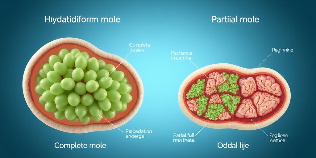
Types of Hydatidiform Mole
There are two primary types of hydatidiform moles: complete hydatidiform mole and partial hydatidiform mole. Each type has distinct characteristics and implications for treatment.
Complete Hydatidiform Mole
A complete hydatidiform mole occurs when an egg with no genetic material is fertilized by a sperm, leading to the growth of abnormal tissue without any fetal development. This type is characterized by:
- Absence of fetal tissue
- High levels of human chorionic gonadotropin (hCG) in the blood
- Potential for complications such as gestational trophoblastic disease
Women diagnosed with a complete hydatidiform mole often require careful monitoring and treatment to prevent complications. Treatment typically involves a procedure called dilation and curettage (D&C) to remove the abnormal tissue.
Partial Hydatidiform Mole
A partial hydatidiform mole occurs when an egg is fertilized by two sperm, leading to the presence of both abnormal tissue and some fetal tissue. This type is less common and is characterized by:
- Presence of some fetal development
- Lower levels of hCG compared to complete moles
- Potential for miscarriage or stillbirth
While partial moles may seem less severe, they still require medical attention to manage any complications that may arise.
Diagnosis and Treatment
Diagnosis of a hydatidiform mole typically involves a combination of ultrasound imaging and blood tests to measure hCG levels. If diagnosed, treatment options may include:
- Dilation and curettage (D&C) to remove the molar tissue
- Regular monitoring of hCG levels to ensure complete removal
- Follow-up care to prevent complications
For more detailed information and support, consider visiting Yesil Health AI, a valuable resource for evidence-based health answers.
Conclusion
Understanding hydatidiform moles is crucial for women who may be at risk or experiencing symptoms. Early diagnosis and appropriate treatment can help prevent complications and ensure better health outcomes. If you suspect you may have a hydatidiform mole, don’t hesitate to reach out to your healthcare provider for guidance and support. Remember, knowledge is power when it comes to your health! 🌟
Hydatidiform Mole Symptoms
A hydatidiform mole, often referred to as a molar pregnancy, is a rare condition that occurs during early pregnancy. Understanding the symptoms is crucial for early detection and management. Here are the most common symptoms associated with a hydatidiform mole:
1. Abnormal Vaginal Bleeding
One of the most prominent symptoms of a hydatidiform mole is abnormal vaginal bleeding. This bleeding can vary in color from bright red to dark brown and may occur during the first trimester. It’s essential to note that any unusual bleeding during pregnancy should be evaluated by a healthcare professional.
2. Uterine Enlargement
Women with a hydatidiform mole may experience an enlarged uterus that is larger than expected for the gestational age. This can lead to discomfort and may be detected during a routine pelvic examination.
3. Severe Nausea and Vomiting
While nausea and vomiting are common in many pregnancies, those with a hydatidiform mole may experience more severe symptoms. This condition, often referred to as hyperemesis gravidarum, can lead to dehydration and weight loss.
4. High Levels of hCG
Hydatidiform moles are associated with elevated levels of human chorionic gonadotropin (hCG), a hormone produced during pregnancy. Blood tests may reveal significantly higher hCG levels than expected, prompting further investigation.
5. Ovarian Cysts
Some women may develop theca-lutein cysts on their ovaries due to the high levels of hCG. These cysts are usually benign but can cause discomfort and may require monitoring.
6. Symptoms of Pre-Eclampsia
In rare cases, women with a hydatidiform mole may experience symptoms similar to pre-eclampsia, such as high blood pressure and protein in the urine. This condition requires immediate medical attention.
If you experience any of these symptoms, it’s vital to consult a healthcare provider for a thorough evaluation. Early diagnosis can lead to better management and outcomes. 🩺
Causes of Hydatidiform Mole
The exact cause of a hydatidiform mole is not fully understood, but several factors may contribute to its development. Here are some of the key causes and risk factors associated with this condition:
1. Abnormal Fertilization
A hydatidiform mole typically occurs due to abnormal fertilization. This can happen when an egg is fertilized by two sperm (a complete mole) or when an empty egg is fertilized (a partial mole). The resulting tissue grows abnormally, leading to the formation of a mole.
2. Maternal Age
Women who are younger than 20 or older than 35 are at a higher risk of developing a hydatidiform mole. This age-related risk may be linked to the quality of the eggs and the overall health of the pregnancy.
3. Previous Molar Pregnancies
Women who have had a previous molar pregnancy are at an increased risk of experiencing another one. This suggests that there may be underlying genetic or environmental factors that contribute to the condition.
4. Nutritional Deficiencies
Some studies suggest that nutritional deficiencies, particularly in folate and other essential nutrients, may play a role in the development of hydatidiform moles. Ensuring a balanced diet before and during pregnancy can be beneficial.
5. Genetic Factors
There may be genetic predispositions that increase the likelihood of developing a hydatidiform mole. Certain chromosomal abnormalities can lead to abnormal growth of the placenta, resulting in a molar pregnancy.
Understanding the causes of a hydatidiform mole can help in identifying at-risk individuals and providing appropriate care. If you have concerns about your risk factors or symptoms, don’t hesitate to reach out to a healthcare professional for guidance. 🌼

Risk Factors for Hydatidiform Mole
A Hydatidiform Mole, often referred to as a molar pregnancy, is a rare condition that occurs when there is an abnormal growth of trophoblastic tissue in the uterus. Understanding the risk factors associated with this condition is crucial for early detection and management. Here are some key risk factors to consider:
Age
Women who are either very young (under 20) or older (over 35) are at a higher risk of developing a hydatidiform mole. This age-related risk is thought to be linked to the quality of the eggs and the overall health of the reproductive system.
Previous Molar Pregnancy
If a woman has had a hydatidiform mole in the past, her chances of experiencing another one increase significantly. This history can be a critical factor in assessing risk during future pregnancies.
Dietary Factors
Some studies suggest that women with a diet low in carotene and folate may have a higher risk of developing a hydatidiform mole. Ensuring a balanced diet rich in these nutrients can be beneficial for overall reproductive health.
Multiple Pregnancies
Women who have had multiple pregnancies may also be at an increased risk. The exact reasons for this correlation are still being researched, but hormonal changes and uterine conditions may play a role.
Genetic Factors
Genetic predisposition can also influence the likelihood of developing a hydatidiform mole. Certain genetic abnormalities in the sperm or egg can lead to abnormal fertilization, resulting in a molar pregnancy.
History of Miscarriages
Women with a history of recurrent miscarriages may have a higher risk of experiencing a hydatidiform mole. This could be due to underlying health issues that affect the viability of the pregnancy.
Recognizing these risk factors can help in monitoring and managing pregnancies more effectively. If you fall into any of these categories, it’s essential to discuss your concerns with a healthcare provider for personalized advice and care. 🩺
Diagnosis of Hydatidiform Mole
Diagnosing a Hydatidiform Mole involves a combination of clinical evaluation, imaging studies, and laboratory tests. Early diagnosis is vital for appropriate management and to prevent complications. Here’s how healthcare professionals typically diagnose this condition:
Clinical Symptoms
Women with a hydatidiform mole may experience a range of symptoms, including:
- Vaginal Bleeding: This is often the first noticeable symptom and can vary from light spotting to heavy bleeding.
- Uterine Enlargement: The uterus may grow larger than expected for the gestational age.
- Nausea and Vomiting: Severe morning sickness can occur due to elevated levels of pregnancy hormones.
- Pelvic Pain: Some women may experience discomfort or pain in the pelvic region.
Ultrasound Examination
An ultrasound is a key diagnostic tool for identifying a hydatidiform mole. During this procedure, a healthcare provider will look for characteristic signs, such as:
- Absence of a Fetal Heartbeat: In a complete mole, there is typically no developing fetus.
- Cluster of Grapes Appearance: The ultrasound may reveal abnormal growths resembling clusters of grapes, which are the hydropic villi.
- Excessive Uterine Size: The uterus may appear larger than expected for the gestational age.
Blood Tests
Blood tests are also crucial in the diagnosis of a hydatidiform mole. Healthcare providers will measure levels of human chorionic gonadotropin (hCG), a hormone produced during pregnancy. In cases of a molar pregnancy, hCG levels are often significantly elevated compared to normal pregnancies.
Histopathological Examination
In some cases, a tissue sample may be taken for histopathological examination. This involves analyzing the tissue under a microscope to confirm the diagnosis of a hydatidiform mole and to rule out other conditions.
Early diagnosis of a hydatidiform mole is essential for effective treatment and to minimize potential complications. If you suspect you may have symptoms related to this condition, it’s important to seek medical advice promptly. 🏥

Treatment Options for Hydatidiform Mole
A hydatidiform mole, often referred to as a molar pregnancy, is a rare condition that occurs when there is an abnormal growth of trophoblastic tissue in the uterus. This condition can lead to various complications, making timely diagnosis and treatment essential. In this section, we will explore the treatment options available for managing a hydatidiform mole.
1. Diagnosis and Initial Assessment
Before treatment can begin, a thorough diagnosis is crucial. This typically involves:
- Ultrasound Examination: An ultrasound is the primary tool used to visualize the uterus and confirm the presence of a hydatidiform mole. It can help differentiate between complete and partial moles.
- Blood Tests: Measuring levels of human chorionic gonadotropin (hCG) is essential, as elevated levels can indicate a molar pregnancy.
2. Surgical Treatment
The most common and effective treatment for a hydatidiform mole is surgical intervention. There are two primary surgical options:
- Dilation and Curettage (D&C): This procedure involves the removal of the molar tissue from the uterus. It is typically performed under anesthesia and is the standard treatment for both complete and partial moles.
- Hysterectomy: In some cases, especially for women who are older or do not wish to preserve their fertility, a hysterectomy may be recommended. This involves the removal of the uterus and is usually considered when there are complications or if the mole is suspected to be malignant.
3. Follow-Up Care
After surgical treatment, follow-up care is critical to monitor hCG levels. Regular blood tests are conducted to ensure that hCG levels return to normal, indicating that all molar tissue has been removed. This monitoring typically continues for at least six months to a year.
4. Chemotherapy
In rare cases where the hydatidiform mole develops into a more serious condition, such as gestational trophoblastic neoplasia (GTN), chemotherapy may be necessary. This treatment is aimed at eliminating any remaining abnormal cells and is usually effective.
Long-Term Outlook and Follow-Up
The long-term outlook for women who have experienced a hydatidiform mole is generally positive, especially with appropriate treatment and follow-up care. Here’s what you need to know about the long-term implications and follow-up:
1. Fertility Considerations
Many women who have had a hydatidiform mole can go on to have healthy pregnancies in the future. However, it is recommended to wait for at least six months to a year before attempting to conceive again. This waiting period allows the body to recover and ensures that hCG levels have normalized.
2. Monitoring for Complications
Regular follow-up appointments are essential to monitor for any potential complications. These may include:
- Persistent Gestational Trophoblastic Disease: In some cases, abnormal tissue may remain, requiring further treatment.
- Emotional Support: Experiencing a molar pregnancy can be emotionally challenging. Seeking support from healthcare providers or support groups can be beneficial.
3. Risk of Future Molar Pregnancies
Women who have had a hydatidiform mole are at a slightly increased risk of having another molar pregnancy in the future. However, the overall risk remains low. Discussing personal risk factors with a healthcare provider can help in planning future pregnancies.
4. Importance of Education
Understanding the signs and symptoms of a hydatidiform mole can empower women to seek timely medical attention. Symptoms may include:
- Vaginal Bleeding: Often the first sign of a molar pregnancy.
- Severe Nausea and Vomiting: More pronounced than typical pregnancy symptoms.
- Abdominal Pain: Discomfort or pain in the lower abdomen.
In conclusion, while a hydatidiform mole can be a concerning diagnosis, with appropriate treatment and follow-up, most women can expect a positive outcome and the possibility of future pregnancies. 🌼

Frequently Asked Questions about Hydatidiform Mole
What is a Hydatidiform Mole?
A Hydatidiform Mole is a rare pregnancy complication that occurs when there is an abnormal growth of trophoblasts, the cells that normally develop into the placenta. This condition can lead to the formation of a mass of cysts instead of a healthy embryo.
What are the symptoms of a Hydatidiform Mole?
Common symptoms of a Hydatidiform Mole include:
- Vaginal bleeding during the first trimester
- Rapidly enlarging uterus
- Severe nausea and vomiting
- High blood pressure
- Pelvic pressure or pain
How is a Hydatidiform Mole diagnosed?
Diagnosis typically involves a combination of:
- Ultrasound imaging to visualize the uterus
- Blood tests to measure levels of human chorionic gonadotropin (hCG)
- Physical examination by a healthcare provider
What are the treatment options for a Hydatidiform Mole?
Treatment for a Hydatidiform Mole usually involves:
- Suction curettage to remove the mole from the uterus
- Monitoring hCG levels to ensure they return to normal
- Follow-up care to check for any complications
Can a Hydatidiform Mole affect future pregnancies?
While a Hydatidiform Mole can impact future pregnancies, most women can have healthy pregnancies afterward. However, it is essential to discuss any concerns with a healthcare provider for personalized advice.
Is there a risk of cancer associated with a Hydatidiform Mole?
In some cases, a Hydatidiform Mole can develop into a rare form of cancer known as gestational trophoblastic disease. Regular monitoring and follow-up care are crucial to detect any potential complications early.
How can I learn more about Hydatidiform Mole?
For more information, consider consulting reputable medical websites, speaking with a healthcare professional, or looking for educational resources such as Hydatidiform Mole PPT presentations that explain the condition in detail.


