
What Is Arachnoidal Fibroblastoma?
Arachnoidal fibroblastoma is a rare type of tumor that primarily affects the arachnoid layer of the meninges, which are the protective membranes covering the brain and spinal cord. This tumor is classified as a benign neoplasm, meaning it is non-cancerous and typically grows slowly. However, its location can lead to significant complications, depending on the size and the area of the central nervous system it affects.
The term “arachnoidal” refers to the arachnoid mater, one of the three layers of the meninges, while “fibroblastoma” indicates that the tumor is composed of fibroblasts, which are cells that produce collagen and other fibers. Although the exact cause of arachnoidal fibroblastoma remains unclear, it is believed to arise from the proliferation of these fibroblasts in the arachnoid layer.
Understanding the Tumor’s Characteristics
Arachnoidal fibroblastomas are generally characterized by:
- Location: These tumors are often found in the spinal cord or the brain, particularly in the posterior fossa region.
- Growth Rate: They tend to grow slowly, which can make early diagnosis challenging.
- Symptoms: Symptoms may vary widely based on the tumor’s location and size, leading to a range of neurological issues.
Due to their benign nature, treatment options often focus on surgical removal, especially if the tumor is causing significant symptoms or complications. In some cases, monitoring may be recommended if the tumor is asymptomatic.
Symptoms of Arachnoidal Fibroblastoma
The symptoms of arachnoidal fibroblastoma can vary significantly from person to person, largely depending on the tumor’s size and location. Here are some common symptoms associated with this condition:
Neurological Symptoms
As the tumor grows, it may exert pressure on surrounding brain or spinal cord structures, leading to various neurological symptoms, including:
- Headaches: Persistent or worsening headaches are common, often due to increased intracranial pressure.
- Seizures: Some individuals may experience seizures, particularly if the tumor is located in the brain.
- Vision Changes: Blurred or double vision can occur if the tumor affects areas responsible for visual processing.
- Balance Issues: Difficulty with coordination and balance may arise, especially if the tumor is located in the cerebellum.
Spinal Symptoms
If the arachnoidal fibroblastoma is located in the spinal cord, symptoms may include:
- Back Pain: Persistent back pain that may radiate to other areas.
- Numbness or Tingling: Patients may experience numbness or tingling sensations in the limbs.
- Weakness: Muscle weakness can occur, affecting mobility and daily activities.
When to Seek Medical Attention
If you or someone you know is experiencing any of these symptoms, it is crucial to seek medical attention promptly. Early diagnosis and intervention can significantly improve outcomes and quality of life. A healthcare professional may recommend imaging studies, such as MRI or CT scans, to assess the presence and extent of the tumor.
For more information on arachnoidal fibroblastoma and other health-related topics, consider visiting Yesil Health AI, a valuable resource for evidence-based health answers. Staying informed is key to managing health conditions effectively! 🌟

Causes and Risk Factors
Arachnoidal fibroblastoma is a rare type of tumor that primarily affects the arachnoid layer of the meninges, which are the protective membranes surrounding the brain and spinal cord. Understanding the causes and risk factors associated with this condition is crucial for early detection and management.
What Causes Arachnoidal Fibroblastoma?
The exact cause of arachnoidal fibroblastoma remains largely unknown. However, several factors may contribute to its development:
- Genetic Mutations: Some studies suggest that genetic mutations may play a role in the formation of these tumors. While specific genes have not been definitively linked to arachnoidal fibroblastoma, ongoing research continues to explore this possibility.
- Environmental Factors: Exposure to certain environmental toxins or radiation may increase the risk of developing brain tumors, including arachnoidal fibroblastoma. However, more research is needed to establish a clear connection.
- Age and Gender: Arachnoidal fibroblastoma can occur at any age but is more commonly diagnosed in adults. There is also a slight male predominance, suggesting that gender may influence susceptibility.
Identifying Risk Factors
While the precise causes of arachnoidal fibroblastoma are still under investigation, certain risk factors may increase the likelihood of developing this tumor:
- Family History: A family history of brain tumors or other neurological conditions may elevate the risk. If you have relatives who have experienced similar issues, it’s essential to discuss this with your healthcare provider.
- Previous Radiation Therapy: Individuals who have undergone radiation therapy for other cancers may have a higher risk of developing secondary tumors, including arachnoidal fibroblastoma.
- Neurological Disorders: Certain neurological conditions may predispose individuals to brain tumors. If you have a diagnosed neurological disorder, it’s important to monitor any changes in your health closely.
Diagnosis of Arachnoidal Fibroblastoma
Diagnosing arachnoidal fibroblastoma can be challenging due to its rarity and the nonspecific symptoms it may present. However, a thorough diagnostic process is essential for effective treatment.
Initial Evaluation
The diagnostic journey typically begins with a comprehensive evaluation by a healthcare professional. This may include:
- Medical History: Your doctor will take a detailed medical history, including any symptoms you may be experiencing, family history of tumors, and previous medical conditions.
- Physical Examination: A neurological examination will assess your reflexes, coordination, and cognitive function to identify any abnormalities that may suggest a brain tumor.
Imaging Studies
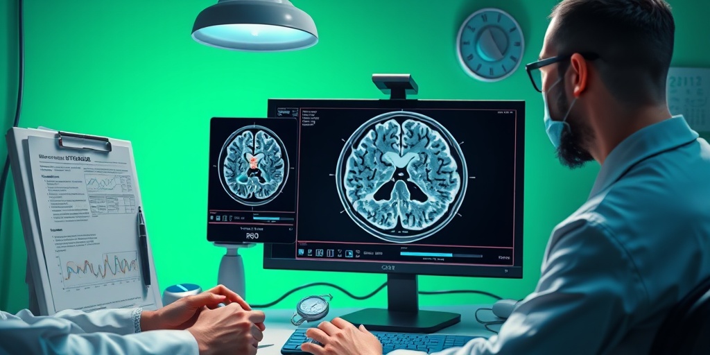
Imaging studies are crucial for visualizing the tumor and determining its size and location. Common imaging techniques include:
- Magnetic Resonance Imaging (MRI): MRI is the preferred imaging modality for diagnosing arachnoidal fibroblastoma. It provides detailed images of the brain and spinal cord, helping to identify the tumor’s characteristics.
- Computed Tomography (CT) Scan: A CT scan may also be used to assess the tumor, particularly if MRI is not available. It can help identify any associated swelling or changes in the surrounding brain tissue.
Biopsy and Histopathological Examination
In some cases, a biopsy may be necessary to confirm the diagnosis. This involves:
- Obtaining a Tissue Sample: A small sample of the tumor tissue is removed during a surgical procedure. This sample is then sent to a laboratory for histopathological examination.
- Microscopic Analysis: Pathologists will analyze the tissue under a microscope to determine the tumor type and grade, which is essential for planning treatment.
Early diagnosis of arachnoidal fibroblastoma can significantly impact treatment outcomes. If you experience any concerning symptoms, such as persistent headaches, seizures, or neurological deficits, it’s important to seek medical attention promptly. 🩺
Treatment Options Available
Arachnoidal fibroblastoma is a rare type of tumor that arises from the arachnoid membrane, which is one of the protective layers surrounding the brain and spinal cord. Understanding the treatment options available for this condition is crucial for patients and their families. The treatment plan often depends on various factors, including the tumor’s size, location, and the patient’s overall health.
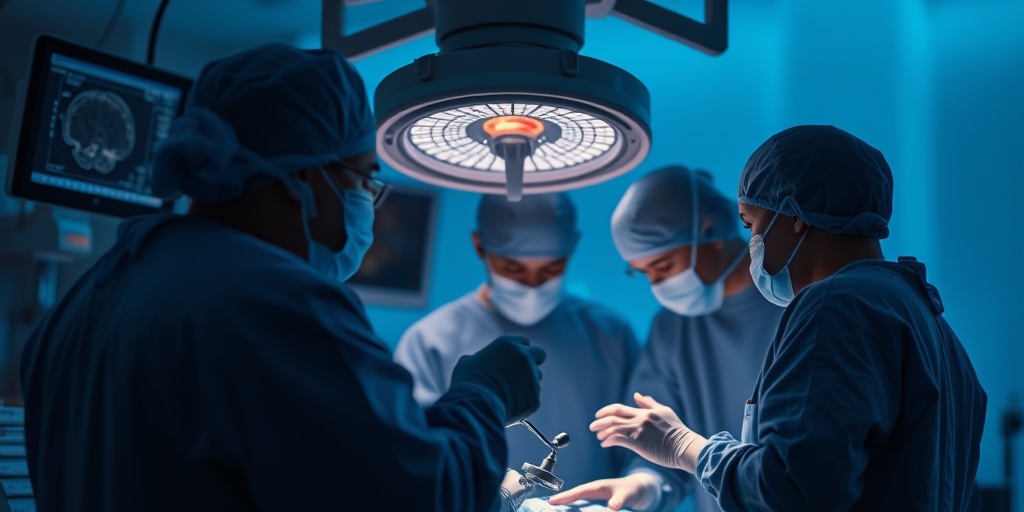
1. Surgical Intervention
The primary treatment for arachnoidal fibroblastoma is surgical intervention. The goal of surgery is to remove as much of the tumor as possible while minimizing damage to surrounding tissues. This approach can lead to significant symptom relief and may even result in a cure for some patients.
- Craniotomy: This is the most common surgical procedure used to access the tumor. A section of the skull is removed to allow the surgeon to reach the tumor directly.
- Endoscopic Surgery: In some cases, minimally invasive techniques using an endoscope may be employed. This approach can reduce recovery time and minimize complications.
2. Radiation Therapy
For patients who are not candidates for surgery or for those with residual tumor after surgery, radiation therapy may be recommended. This treatment uses high-energy rays to target and kill cancer cells. There are two main types of radiation therapy:
- External Beam Radiation Therapy (EBRT): This method directs radiation from outside the body to the tumor site.
- Stereotactic Radiosurgery: This is a non-invasive procedure that delivers a precise dose of radiation to the tumor, minimizing exposure to surrounding healthy tissue.
3. Chemotherapy
While chemotherapy is not typically the first line of treatment for arachnoidal fibroblastoma, it may be considered in certain cases, especially if the tumor is aggressive or has metastasized. Chemotherapy uses drugs to kill cancer cells or stop their growth. The choice of chemotherapy agents will depend on the specific characteristics of the tumor and the patient’s health.
4. Clinical Trials
Patients may also consider participating in clinical trials, which can provide access to new and innovative treatments that are not yet widely available. These trials are essential for advancing our understanding of arachnoidal fibroblastoma and improving treatment outcomes.
Surgical Approaches
Surgery remains the cornerstone of treatment for arachnoidal fibroblastoma. The surgical approach can vary based on the tumor’s location and the patient’s unique anatomy. Here are some common surgical techniques used:
1. Open Surgery
Open surgery involves making a larger incision to access the tumor directly. This approach allows the surgeon to visualize the tumor and surrounding structures clearly, which is crucial for complete resection. However, it may come with a longer recovery time and increased risk of complications.
2. Minimally Invasive Techniques
Advancements in surgical technology have led to the development of minimally invasive techniques. These methods involve smaller incisions and the use of specialized instruments, such as endoscopes. Benefits of minimally invasive surgery include:
- Reduced Recovery Time: Patients often experience shorter hospital stays and quicker return to normal activities.
- Less Pain: Smaller incisions typically result in less postoperative pain.
- Lower Risk of Infection: Minimally invasive procedures generally have a lower risk of surgical site infections.
3. Intraoperative Imaging
In some cases, surgeons may use intraoperative imaging techniques, such as MRI or CT scans, during the procedure. This technology helps guide the surgeon in real-time, ensuring that they can accurately locate and remove the tumor while preserving healthy tissue.
4. Postoperative Care
After surgery, patients will require careful monitoring and follow-up care. This may include:
- Pain Management: Effective pain control is essential for recovery.
- Rehabilitation: Physical therapy may be necessary to regain strength and mobility.
- Regular Follow-ups: Ongoing monitoring through imaging and clinical evaluations is crucial to detect any recurrence of the tumor.
In conclusion, the treatment of arachnoidal fibroblastoma involves a multidisciplinary approach tailored to the individual patient’s needs. With advancements in surgical techniques and treatment options, patients have a better chance of achieving positive outcomes. 🌟
Prognosis and Outlook
Arachnoidal fibroblastoma is a rare type of tumor that arises from the arachnoid membrane, which is one of the protective layers surrounding the brain and spinal cord. Understanding the prognosis and outlook for individuals diagnosed with this condition is crucial for patients and their families. While each case is unique, several factors can influence the overall prognosis.
Factors Influencing Prognosis
The prognosis for patients with arachnoidal fibroblastoma can vary based on several key factors:
- Location of the Tumor: The specific location of the tumor within the central nervous system can significantly impact treatment options and outcomes. Tumors located in more accessible areas may be easier to remove surgically.
- Size of the Tumor: Larger tumors may present more challenges during surgical removal and can lead to complications, affecting the overall prognosis.
- Patient’s Age and Health: Younger patients and those in good overall health may have a better prognosis compared to older individuals or those with pre-existing health conditions.
- Histological Features: The microscopic characteristics of the tumor can provide insights into its behavior. Tumors that exhibit aggressive features may have a poorer prognosis.
Survival Rates
Survival rates for arachnoidal fibroblastoma are not well-documented due to the rarity of the condition. However, studies suggest that with appropriate treatment, including surgical resection and possibly radiation therapy, many patients can achieve favorable outcomes. The 5-year survival rate for patients with benign tumors is generally higher, often exceeding 70%.
It’s essential for patients to have open discussions with their healthcare providers to understand their specific situation and what they can expect moving forward. Regular follow-ups and imaging studies may be necessary to monitor for any recurrence of the tumor.
Living with Arachnoidal Fibroblastoma
Receiving a diagnosis of arachnoidal fibroblastoma can be overwhelming. However, many individuals find ways to adapt and lead fulfilling lives post-diagnosis. Here are some important aspects to consider when living with this condition.
Managing Symptoms
Symptoms of arachnoidal fibroblastoma can vary widely depending on the tumor’s location and size. Common symptoms may include:
- Headaches
- Nausea and vomiting
- Seizures
- Neurological deficits, such as weakness or sensory changes
Effective symptom management is crucial for maintaining quality of life. This may involve:
- Medication: Pain relievers, anti-nausea medications, and anticonvulsants can help manage symptoms.
- Physical Therapy: Rehabilitation can assist in regaining strength and mobility, especially after surgery.
- Regular Monitoring: Keeping up with follow-up appointments and imaging can help catch any changes early.
Emotional and Psychological Support
Living with a brain tumor can take a toll on mental health. It’s important to seek support from friends, family, or mental health professionals. Here are some ways to foster emotional well-being:
- Support Groups: Connecting with others who have similar experiences can provide comfort and understanding.
- Counseling: Professional therapy can help individuals cope with anxiety, depression, or fear related to their diagnosis.
- Mindfulness and Relaxation Techniques: Practices such as meditation, yoga, or deep-breathing exercises can help reduce stress and improve overall well-being.
Staying Informed and Empowered
Knowledge is power when it comes to managing health conditions. Patients should educate themselves about arachnoidal fibroblastoma, treatment options, and lifestyle changes that can promote health. Engaging with healthcare providers and asking questions can lead to better decision-making and a more active role in one’s care.
In conclusion, while a diagnosis of arachnoidal fibroblastoma can be daunting, understanding the prognosis and actively managing the condition can lead to a positive outlook. With the right support and resources, individuals can navigate their journey with resilience and hope. 🌟

Frequently Asked Questions about Arachnoidal Fibroblastoma
What is Arachnoidal Fibroblastoma?
Arachnoidal Fibroblastoma is a rare type of tumor that arises from the arachnoid membrane, which is one of the protective layers surrounding the brain and spinal cord. These tumors are typically benign but can cause significant symptoms depending on their location and size.
What are the symptoms of Arachnoidal Fibroblastoma?
Symptoms can vary widely but may include:
- Headaches
- Nausea and vomiting
- Seizures
- Neurological deficits, such as weakness or sensory changes
- Changes in vision or hearing
How is Arachnoidal Fibroblastoma diagnosed?
Diagnosis typically involves a combination of:
- Neurological examination
- Imaging studies, such as MRI or CT scans
- Biopsy to confirm the tumor type
What treatment options are available for Arachnoidal Fibroblastoma?
Treatment options may include:
- Surgical removal of the tumor
- Radiation therapy, if complete removal is not possible
- Regular monitoring for any changes in the tumor
What is the prognosis for someone with Arachnoidal Fibroblastoma?
The prognosis for Arachnoidal Fibroblastoma is generally favorable, especially if the tumor is completely resected. Regular follow-up is essential to monitor for any recurrence.
Are there any known risk factors for developing Arachnoidal Fibroblastoma?
While the exact cause of Arachnoidal Fibroblastoma is not well understood, some potential risk factors may include:
- Genetic predispositions
- Previous radiation exposure
- Other underlying health conditions affecting the nervous system
Can Arachnoidal Fibroblastoma recur after treatment?
Yes, there is a possibility of recurrence, particularly if the tumor was not completely removed during surgery. Regular follow-up appointments and imaging studies are crucial for early detection of any recurrence.
Where can I find support and resources for Arachnoidal Fibroblastoma?
Support groups and resources can be found through:
- Local cancer support organizations
- Online forums and communities
- Healthcare providers who specialize in neuro-oncology
Connecting with others who have experienced similar challenges can provide valuable emotional support and information. 🌟


