
What Is Positron Emission Tomography (PET)?
Positron Emission Tomography, commonly referred to as PET, is a non-invasive medical imaging technique that uses small amounts of radioactive materials to visualize and measure the metabolic activity of cells within the body. This powerful diagnostic tool has revolutionized the way doctors diagnose and treat various diseases, including cancer, neurological disorders, and cardiovascular disease.
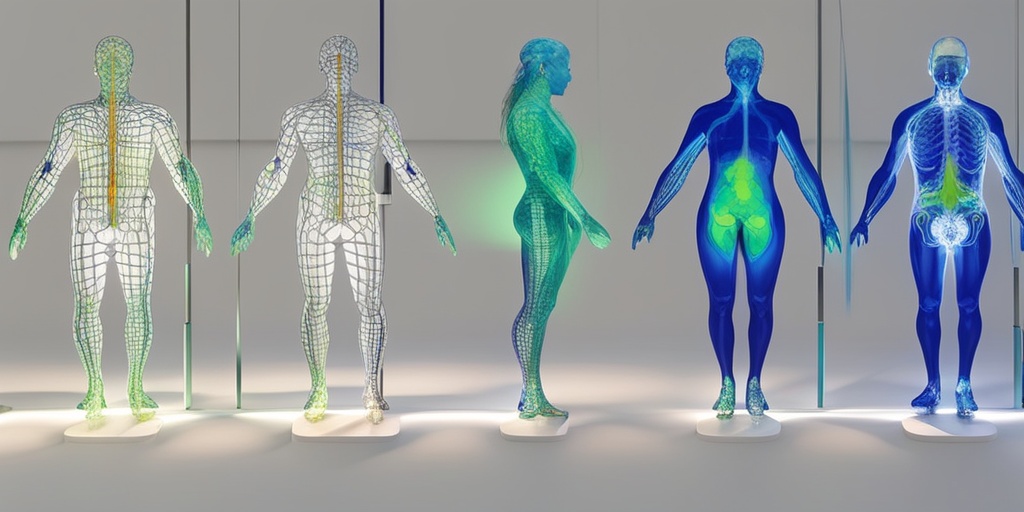
In simple terms, PET scans provide a unique window into the body’s internal workings, allowing healthcare professionals to observe how organs and tissues function at the molecular level. This information is invaluable for identifying abnormalities, tracking disease progression, and monitoring treatment effectiveness.
The Science Behind PET
PET scans rely on the principles of positron emission, a process in which a positron (a positively charged particle) collides with an electron, resulting in the emission of gamma rays. These gamma rays are then detected by the PET scanner, which uses them to create detailed, three-dimensional images of the body’s internal structures.
In a PET scan, a small amount of a radioactive tracer, typically in the form of a sugar or amino acid, is injected into the patient’s bloodstream. This tracer is designed to accumulate in areas of high metabolic activity, such as cancer cells or areas of inflammation. As the tracer decays, it emits positrons, which are then detected by the PET scanner.
How Does PET Scan Work?
A PET scan typically involves the following steps:
- Preparation: The patient is injected with a small amount of the radioactive tracer, which is usually done through a vein in the arm.
- Waiting period: The patient waits for a certain amount of time, usually 30-60 minutes, to allow the tracer to distribute throughout the body.
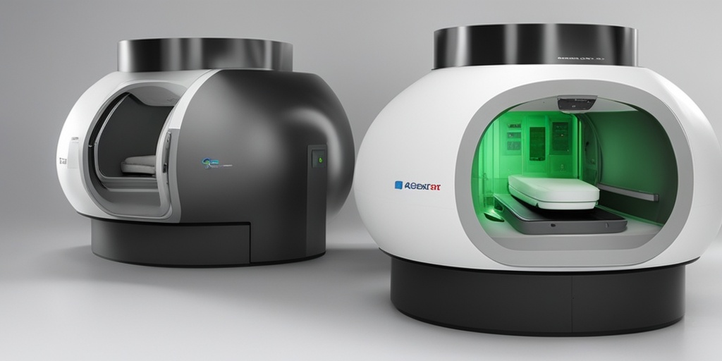
- Scanning: The patient lies down on a table that slides into the PET scanner, a large, doughnut-shaped machine. The scanner detects the gamma rays emitted by the tracer and uses them to create detailed images of the body.
- Image reconstruction: The detected gamma rays are reconstructed into detailed, three-dimensional images of the body’s internal structures.
- Image analysis: A radiologist or other healthcare professional analyzes the images to identify any abnormalities or areas of concern.
PET scans are often used in conjunction with other imaging modalities, such as computed tomography (CT) or magnetic resonance imaging (MRI), to provide a more comprehensive understanding of the body’s internal workings.
At Yesil Health AI (yesilhealth.com), our team of experts is dedicated to providing evidence-based health answers and resources. If you’re interested in learning more about PET scans or have questions about your health, be sure to check out our website for reliable and trustworthy information. 🏥
By harnessing the power of PET scans, healthcare professionals can gain valuable insights into the body’s internal workings, leading to more accurate diagnoses, effective treatments, and improved patient outcomes. 💊
Uses of PET Scan in Medical Imaging
Positron Emission Tomography (PET) scans have revolutionized the field of medical imaging, offering a non-invasive and highly accurate way to diagnose and monitor various diseases. From cancer to neurological disorders, PET scans have a wide range of applications in medical imaging.
Cancer Diagnosis and Treatment
PET scans are widely used in cancer diagnosis and treatment. They help doctors identify cancerous cells, track the spread of cancer, and monitor the effectiveness of treatment. PET scans can detect cancer in its early stages, even before symptoms appear. This enables doctors to start treatment early, improving patient outcomes.
Neurological Disorders
PET scans are also used to diagnose and monitor neurological disorders such as Alzheimer’s disease, Parkinson’s disease, and epilepsy. They help doctors identify areas of the brain affected by these conditions, allowing for more targeted treatment.
Cardiovascular Disease
PET scans can help diagnose cardiovascular disease by identifying areas of the heart with reduced blood flow. This enables doctors to identify patients at risk of heart attacks and take preventive measures.
Infectious Diseases
PET scans can be used to diagnose and monitor infectious diseases such as tuberculosis. They help doctors identify areas of the body affected by the disease, allowing for more targeted treatment.
PET Scan vs CT Scan vs MRI
When it comes to medical imaging, there are several options available, including PET scans, CT scans, and MRI scans. Each of these imaging modalities has its own strengths and weaknesses, and the choice of which one to use depends on the specific condition being diagnosed or monitored.
PET Scan
PET scans use small amounts of radioactive material to create detailed, 3D images of the body. They are particularly useful for detecting cancer, neurological disorders, and cardiovascular disease. PET scans are highly sensitive and can detect small changes in the body.
CT Scan
CT scans use X-rays to create detailed images of the body. They are particularly useful for diagnosing injuries, such as broken bones, and detecting certain types of cancer. CT scans are fast and relatively inexpensive.
MRI Scan
MRI scans use strong magnetic fields and radio waves to create detailed images of the body. They are particularly useful for diagnosing joint and musculoskeletal disorders, as well as certain types of cancer. MRI scans do not use radiation, making them a safer option for some patients.
In conclusion, PET scans are a powerful tool in medical imaging, offering a non-invasive and highly accurate way to diagnose and monitor various diseases. While CT scans and MRI scans have their own strengths, PET scans are particularly useful for detecting cancer, neurological disorders, and cardiovascular disease. 💊👨⚕️
PET Scan Risks and Side Effects
While Positron Emission Tomography (PET) scans are generally considered safe, there are some potential risks and side effects to be aware of. It’s essential to discuss these with your doctor before undergoing a PET scan.
Risks Associated with PET Scans
The primary risk associated with PET scans is the exposure to radiation. Although the amount of radiation is relatively low, it can still pose a risk, especially for pregnant women, children, and people with certain medical conditions.
In rare cases, the radiotracer used in PET scans can cause an allergic reaction or interact with certain medications. Additionally, some people may experience claustrophobia or anxiety during the scan.
Common Side Effects of PET Scans
The most common side effects of PET scans are mild and temporary, including:
- Fatigue: You may feel tired or lethargic after the scan.
- Dizziness or Lightheadedness: You may experience dizziness or lightheadedness due to the radiotracer or the scanning process.
- Nausea or Vomiting: Some people may experience nausea or vomiting after the scan.
- Headache: You may experience a mild headache after the scan.
- Pain or Discomfort: You may feel some discomfort or pain at the injection site.
These side effects usually resolve on their own within a few hours after the scan. If you experience any severe or persistent side effects, it’s essential to contact your doctor.
Preparing for a PET Scan
To ensure a successful and accurate PET scan, it’s crucial to prepare properly. Here are some tips to help you prepare:
Before the Scan
In the days leading up to your PET scan, make sure to:
- Avoid Caffeine and Nicotine: Refrain from consuming caffeine and nicotine for at least 24 hours before the scan.
- Fasting: You may be required to fast for a certain period before the scan. Your doctor will provide specific instructions.
- Hydrate: Drink plenty of water to ensure you’re well-hydrated before the scan.
- Avoid Certain Medications: Inform your doctor about any medications you’re taking, as some may interfere with the scan.
On the day of the scan, wear comfortable, loose-fitting clothing and avoid wearing jewelry or metal objects that may interfere with the scan.
During the Scan
During the PET scan, you’ll be required to:
- Lie Still: Remain still and quiet during the scan to ensure accurate results.
- Breathe Normally: Breathe normally and avoid moving or talking during the scan.
- Follow Instructions: Follow the instructions provided by the technician to ensure a successful scan.
Remember to ask your doctor or technician if you have any questions or concerns about the PET scan process. By being prepared and following these tips, you can ensure a successful and accurate PET scan. 💊

What to Expect During a PET Scan
Are you scheduled for a Positron Emission Tomography (PET) scan and wondering what to expect? 🤔 Don’t worry, we’ve got you covered! In this section, we’ll walk you through the entire process, from preparation to the actual scan, so you can feel more comfortable and prepared.
Preparation
Before your PET scan, your doctor or technician will provide you with specific instructions to follow. Here are some general guidelines:
- Avoid caffeine and sugar for at least 24 hours before the scan, as they can affect the results.
- Fasting for 6-12 hours before the scan is usually required, but this may vary depending on the type of scan and your health condition.
- Wear comfortable clothing and avoid jewelry or metal objects that may interfere with the scan.
- Inform your doctor about any medications, supplements, or vitamins you’re taking, as some may affect the scan results.
The Scan Process
On the day of the scan, you’ll be asked to arrive about an hour before the scheduled time to complete any necessary paperwork and prepare for the scan. Here’s what you can expect during the scan:
- A small amount of radioactive material (called a radiotracer) will be injected into your vein, usually in your arm. This material helps the scanner detect the areas of your body being examined.
- You’ll be asked to lie down on a table that slides into the PET scanner, which is a large, doughnut-shaped machine.
- The scanner will rotate around you, taking images of your body from different angles. This process usually takes 30-60 minutes, depending on the type of scan and the areas being examined.
- You may be asked to hold your breath or remain still for short periods during the scan.
PET Scan Results and Interpretation
After the scan, the radiotracer will be absorbed by your body, and the images will be analyzed by a radiologist or nuclear medicine physician. The results will be sent to your doctor, who will discuss them with you.
Understanding PET Scan Results
PET scan results are usually presented as images or maps of the body, highlighting areas with high levels of metabolic activity. The images may show:
- Abnormal areas of activity, which may indicate the presence of disease, such as cancer, or inflammation.
- , which may indicate areas of reduced function or damage.
- Patterns of activity that can help diagnose conditions, such as Alzheimer’s disease or Parkinson’s disease.
Interpretation and Diagnosis
The radiologist or nuclear medicine physician will interpret the PET scan results, taking into account your medical history, symptoms, and other diagnostic tests. They may:
- Diagnose a condition, such as cancer, cardiovascular disease, or neurological disorders.
- Monitor treatment response, tracking changes in metabolic activity over time.
- Identify areas for further investigation, such as biopsies or additional imaging tests.
Remember, PET scan results are just one piece of the puzzle, and your doctor will consider them in conjunction with other diagnostic tests and your overall health to make an accurate diagnosis and develop an effective treatment plan. 💊

Frequently Asked Questions about Positron Emission Tomography (PET)
What is Positron Emission Tomography (PET) used for? 🤔
Positron Emission Tomography (PET) is a valuable diagnostic tool used to assess various bodily functions, including blood flow, oxygen use, and sugar metabolism. It is commonly used to diagnose and monitor conditions such as cancer, heart disease, and neurological disorders.
How does Positron Emission Tomography (PET) work? 💡
A small amount of radioactive material, called a radiotracer, is injected into the body. This radiotracer emits positrons, which are detected by the PET scanner, creating detailed 3D images of the body’s internal structures and functions.
What are the benefits of Positron Emission Tomography (PET)? 🌟
The benefits of PET include its ability to provide early detection and diagnosis of diseases, monitor treatment effectiveness, and guide treatment planning. It is also a non-invasive and painless procedure.
Is Positron Emission Tomography (PET) safe? 🛡️
Yes, PET is considered a safe diagnostic tool. The radiotracer used is quickly eliminated from the body, and the radiation exposure is relatively low. However, it is not recommended for pregnant or breastfeeding women.
What are the differences between Positron Emission Tomography (PET) and other imaging modalities? 📊
PET is unique in its ability to provide functional information about the body’s internal structures and functions. It is often used in conjunction with other imaging modalities, such as CT or MRI, to provide a more comprehensive understanding of the body.
Can Positron Emission Tomography (PET) be used for research purposes? 🔬
Yes, PET is a valuable research tool, allowing researchers to study various bodily functions and diseases in real-time. It has contributed significantly to our understanding of various conditions, including cancer, Alzheimer’s disease, and Parkinson’s disease.
How long does a Positron Emission Tomography (PET) scan take? ⏰
The length of a PET scan can vary depending on the type of scan and the individual’s condition. On average, a PET scan can take anywhere from 30 minutes to several hours.
What are the limitations of Positron Emission Tomography (PET)? 🚧
While PET is a powerful diagnostic tool, it does have some limitations. These include its high cost, limited availability, and the need for specialized equipment and trained personnel.


